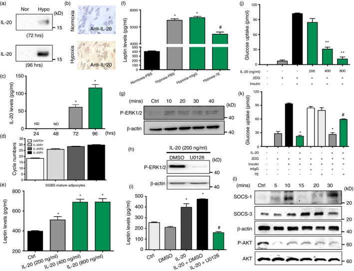
FIGURE 4.

Functions of IL‐20 in SGBS adipocytes. (a) Western blotting of IL‐20 in the conditioned medium under normoxic and hypoxic conditions for the indicated time periods. (b) IHC staining of IL‐20 in mature SGBS cells under hypoxic conditions for 4 days. (c) The level of IL‐20 in mature SGBS cells under hypoxic conditions for 24–96 h was measured using ELISA. *p < 0·05 compared with hypoxia 24 h. ND =nondetectable. (d) The expression of IL‐20R1, IL‐20R2 and IL‐22R1 mRNA in mature SGBS adipocytes was analysed using RT‐qPCR. (e) The level of leptin in IL‐20‐treated SGBS cells (200–800 ng/ml for 14 days in differentiation medium) was measured using ELISA. *p < 0·05 compared with untreated controls. (f) The level of leptin in mIgG‐ or 7E‐treated mature SGBS cells under hypoxic conditions for 96 h was measured using ELISA. *p < 0·05 compared with normoxic controls, #p < 0·05 compared with mIgG‐treated group. (g) Mature SGBS cells were incubated with IL‐20 (200 ng/ml) for the indicated time periods, and then, cell lysates were collected and analysed using immunoblotting with specific antibodies against phospho‐ERK1/2. β‐Actin was an internal control. (h) Western blot of phospho‐ERK1/2 and β‐actin in mature SGBS cells treated with IL‐20 (200 ng/ml) in the presence of the ERK1/2 inhibitor U0126 (10 μM) or vehicle (DMSO). (i) Leptin level in culture supernatants of mature SGBS cells treated with IL‐20 (200 ng/ml) in the presence of the ERK1/2 inhibitor U0126 (10 μM) or vehicle (DMSO). *p < 0·05 compared with untreated controls, #p < 0·05 compared with IL‐20 plus DMSO‐treated group. (j‐k) The differentiated SGBS mature adipocytes were incubated with IL‐20 (200–800 ng/ml), mIgG (8 μg/ml), 7E (8 μg/ml), IL‐20 (800 ng/ml) plus mIgG (8 μg/ml) or IL‐20 (800 ng/ml) plus 7E (8 μg/ml) in serum‐free medium for 24 h, and then, 2‐deoxyglucose uptake was assessed using glucose uptake assay. *p < 0·05, **p < 0·01 compared with untreated controls, #p < 0·05 compared with IL‐20 plus mIgG‐treated group. (l) Western blot of SOCS‐1, SOCS‐3, β‐actin phospho‐AKT and AKT in mature SGBS cells treated with IL‐20 (200 ng/ml) for the indicated time periods. Data in (c), (e), (f), (i), (j) and (k) are expressed as mean ±SEM and are representative of three independent experiments
