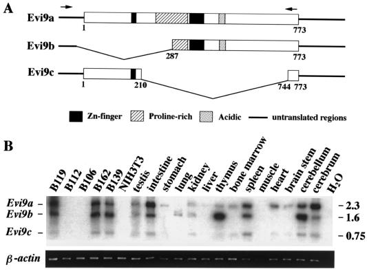
FIG. 3.
Three Evi9 isoforms are expressed in a tissue-specific fashion. (A) Protein structures of three Evi9 isoforms. Arrows indicate the locations of PCR primers used for RT-PCR. (B) Expression patterns of three Evi9 isoforms (upper panel) in various tissues and BXH2 leukemia cell lines were analyzed by RT-PCR. RT-PCR products were gel fractionated, transferred to a nylon membrane, and hybridized with probe AA53. Sizes of PCR products in kilobases are shown on the right. β-Actin (lower panel) was amplified from the same samples to check for RNA quality.

