Figure 6.
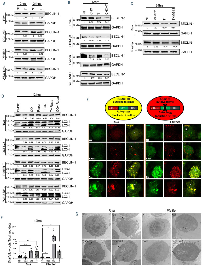
TBL1 modulates cytoprotective autophagy through the SCF complex. (A) Immunoblot showing BECLIN-1 expression in the indicated diffuse large B-cell lymphoma (DLBCL) cell lines after treatment with either dimethylsulfoxide (DMSO) control (NT) or tegavivint (T) for 12 hours and 24 hours (n=3). (B) Immunoblot showing BECLIN-1 expression in the indicated DLBCL cell lines after treatment with either cycloheximide (CHX), tegavivint (T) or the combination (CHX+T) (n=3). (C) Immunoblot showing BECLIN-1 expression in the indicated DLBCL cell lines after treatment with either DMSO control (NT), tegavivint (T), MG132 or the combination (T+MG132) at 24 hours (n=3). (D) Immunoblot showing BECLIN-1 and microtubule-associated protein light-chain 3 (LC3-I and LC3-II) expression in the indicated DLBCL cell lines after treatment with either DMSO control (NT), tegavivint (T), chloroquine (CQ), rapamycin (Rapa) or combinations at 12 hours and 24 hours (n=3). (E) Top: schematic depicting tandem LC3 reporter. Bottom: confocal microscopy images (120x) of the indicated DLBCL cell lines transduced with a lentivirus expressing a tandem LC3 plasmid (mCherry-eGFP-LC3) and subsequently treated with either DMSO control (NT), tegavivint (T), chloroquine (CQ) or rapamycin (Rapa) for 12 hours. (F) Histograms represent quantification of yellow (GFP/mCherry) puncta/total red (mCherry) puncta (%) of no less than 100 cells per condition. n=4, data represent means ± standard error of the mean (SEM). *P<0.05, **P<0.005 by a linear mixed effects model with Holm’s adjustment for multiple comparisons for each cell line. (G) Representative transmission electron microscopy images showing ultrastructural changes in the indicated DLBCL cell lines after treatment with either DMSO control (NT), tegavivint, chloroquine (CQ) or rapamycin (Rapa) at 12 hours. The arrows indicate accumulation of autophagic vacuoles containing cytoplasmic material after exposure to tegavivint or chloroquine (n=3). Chloroquine: 50uM and rapamycin: 10uM for all cell lines.

