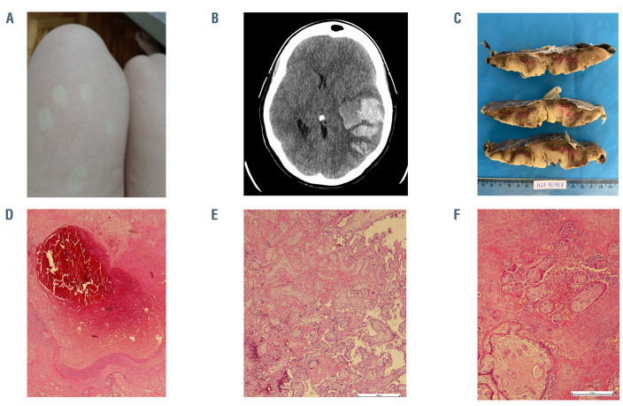
Figure 1.
Pictures of skin rash, cerebral hemorrhage, and placenta. (A) Generalized skin rash on legs with spontaneous resolution; (B) brain computed tomography with large acute intraparenchymal hematoma in the temporal lobe, insula, and temporoparietal transition of the left cerebral hemisphere measuring approximately 10.1 x 5.4 x 5.5 cm surrounded by vasogenic edema with midline shift to the right, herniation of the uncus, marked compressive effect on the midbrain and subtotal collapse of the supratentorial ventricular system; (C) cut surface of the placenta shows pale parenchyma with irregular and firm red-brown areas; (D) occlusive thrombus in a decidual vessel surrounded by a recent infarct; (E) chorionic villi encased by perivillous fibrin and red blood cells with some preservation of the trophoblast layer and villous stroma; villous capillaries are sparse; (F) area with massive intervillous fibrin deposition, some leukocytes and red blood cells with degenerative villous configuring an intervillous thrombosis.

