FIG 5.
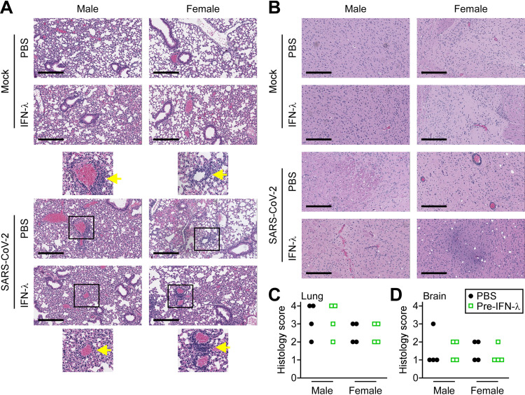
Lung and brain histology following SARS-CoV-2 infection. (A) Representative hematoxylin and eosin (H&E) staining of mouse lung tissue sections. Top panels show images of lungs from uninfected mice (mock) treated with PBS or IFN-λ for 5 days. Bottom panels show representative lung sections from mice that were pretreated with PBS or IFN-λ and infected with 1 × 10³ PFU SARS-CoV-2 for 5 days. Scale bar, 300 μm. Magnified images are shown with arrows identifying perivascular immune cell infiltration. (B) H&E-stained sections of brain tissue from mice treated as in panel A. Quantitative histology scoring for lungs (C) and brains (D) of male and female mice is described in Table S2 in the supplemental material.

