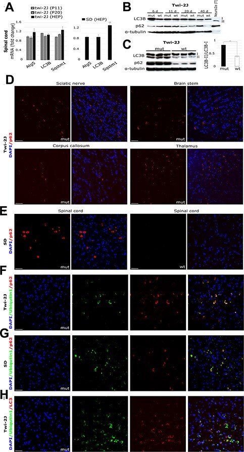
Figure 4 .

Autophagosomal/lysosomal dysfunction accompanies Ripk1 expression in Krabbe and SD. Autophagy was studied by (A) RT-qPCR of total RNA in spinal cord at three stages of disease in twi-2J and at the HEP in SD, with markers Atg5 (Autophagy related 5), LC3B (Microtubule-associated protein 1 light chain 3 beta), and Sqstm1 (Sequestosome 1 codes for p62); (B) Immunoblot of LC3B and p62 in twi-2J spinal cord at different ages. Protein extracts from Neuro2a chloroquine-treated cells were used as positive control of upregulated autophagy; (C) Immunoblot to LC3B and p62 and densitometry in twi-2J brain stem at HEP. Seventy-five microgram protein homogenate was loaded per lane. Blots were stripped of first antibody and re-probed with subsequent antibodies. Fluorescent IHC staining of p62 in twi-2J sciatic nerve and brain (D), and SD spinal cord at HEP (E). Dual IHC staining of ubiquitin1 and p62 in twi-2J spinal cord at HEP (F) and SD (G). (H) Dual IHC staining of ubiquitin1 and LC3B in twi-2J spinal cord at HEP. Nuclear stain: DAPI (4′,6-Diamidine-2′-phenylindole dihydrochloride), mutant (mut), wild type (wt). Student’s t-test; *P ≤ 0.05; **P ≤ 0.01.
