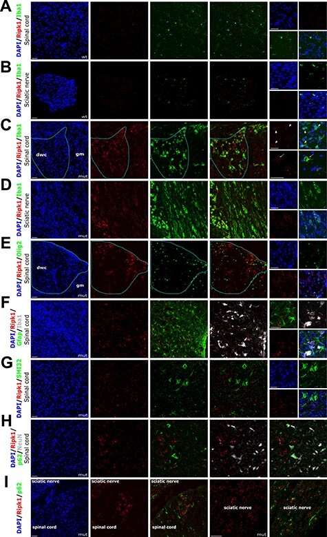
Figure 6 .

Ripk1 localizes to activated macrophage/microglia. (A-I) Combined ISH with RNAscope probe Mm-Ripk1-C1 and fluorescent IHC staining of twi-2J with cell type-specific markers for macrophage/microglia Iba1(A-D), oligodendroglia Olig2 (E), macrophage/microglia Iba1 and astroglia Gfap (F), non-phosphorylated neurofilament protein SMI32 (G), p62 and neuronal marker NeuN (H), and p62 (I). Nuclear stain: DAPI (4′,6-Diamidine-2′-phenylindole dihydrochloride. Mutant (mut), wild type (wt), dorsal white column (dwc), grey matter (gm). Scale bars: 50 μm.
