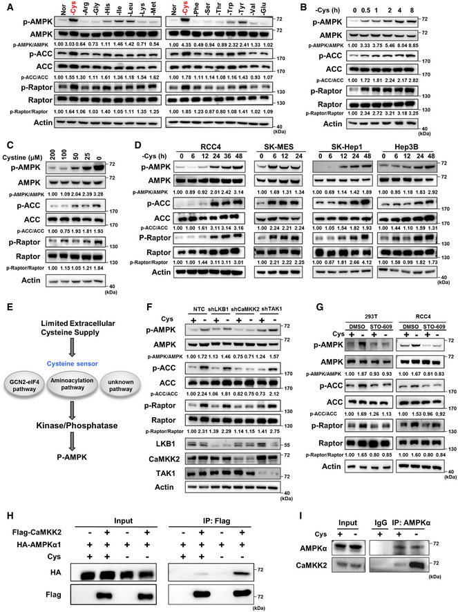
Figure 1. Cystine deprivation activates AMPK through CaMKK2.

-
A293T cells were cultured in complete medium made of dialysed serum for 24 h which was then replaced with medium in which one amino acid was eliminated and cultured for 8 h. Then, WB was used to measure p‐AMPK, p‐ACC, p‐Raptor and total AMPK, ACC, Raptor protein expression. “Nor” = normal medium. Actin served as the loading control. The red text "‐Cys" indicates that cystine deficiency most significantly activates AMPK and its substrates.
-
B–D293T cells were treated with cystine‐deficient medium for 0, 0.5, 1, 2, 4 or 8 h (B) or treated with medium containing 200, 100, 50, 25 or 0 µM cystine for 8 h (C); RCC4, SK‐MES, SK‐hep‐1 or Hep3B cells were treated with cystine‐deficient medium for indicated time (D). Protein levels of p‐AMPK, p‐ACC, p‐Raptor and total AMPK, ACC, Raptor were measured by WB, and actin served as the loading control.
-
EA schematic diagram of the hypothetical mechanism showing how cystine deprivation induces AMPK activation.
-
FWB analysis of p‐AMPK, p‐ACC, p‐Raptor and total AMPK, ACC, Raptor protein in 293T cells transfected with shRNAs targeting LKB1, CaMKK2, TAK1 or a non‐targeting control (NTC) that were treated with cystine‐deficient medium for 8 h. Actin served as the loading control.
-
GWB analysis of p‐AMPK, p‐ACC, p‐Raptor and total AMPK, ACC, Raptor protein in 293T cells and RCC4 cells treated with 1 µg/ml STO‐609 or DMSO for 8 h during cystine deprivation. Actin served as the loading control.
-
H293T cells were transfected with HA‐AMPKα1 alone or together with Flag‐CaMKK2 for 48 h and then cultured with cystine‐deficient medium or complete medium for 8 h. Cell lysates were immunoprecipitated with anti‐Flag, and then, WB analysis was performed.
-
I293T cells cultured with cystine‐deficient medium or complete medium for 8 h were harvested and subjected to immunoprecipitation with anti‐AMPKα, followed by WB analysis with anti‐AMPKα and anti‐CaMKK2.
Source data are available online for this figure.
