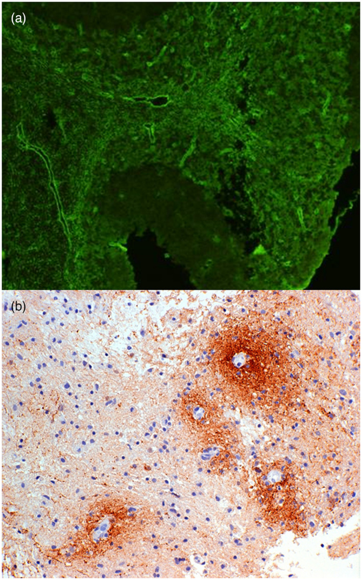
FIGURE 1.

Perivascular distribution of aquaporin‐4 (AQP4) antibodies and complement in neuromyelitis optica spectrum disorder (NMOSD). AQP4 antibody positive immunofluorescence (green) in mouse cerebellum [serum dilution 1:40, goat anti‐human immunoglobulin (Ig)G F(ab)2 fluorescein isothiocyanate, ×200 magnification], showing microvessel staining of the granular layer, molecular layer and white matter (a). Section of early white matter lesion in NMOSD immunostained (brown) for Cd3 showing typical perivascular complement deposition around small blood vessels (b)
