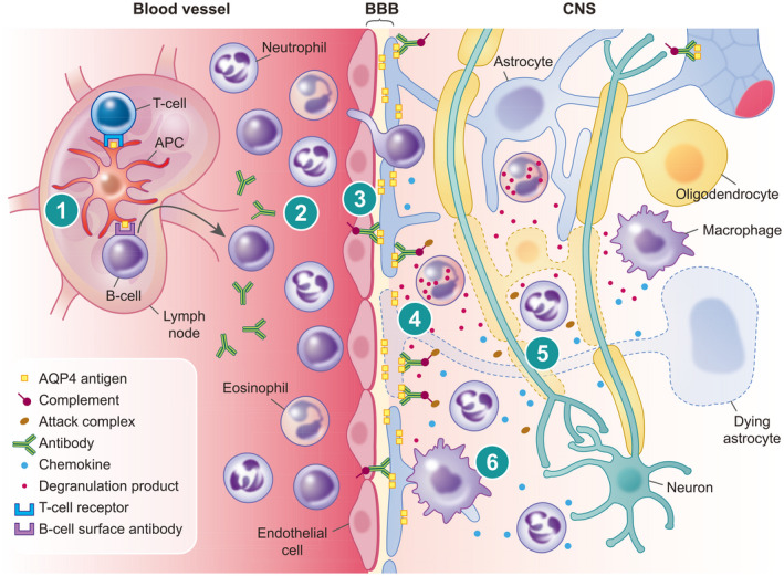
FIGURE 2.

Schematic representation of the pathogenesis of neuromyelitis optica spectrum disorder (NMOSD). (1) Presentation of aquaporin‐4 (AQP4) antigen via an antigen‐presenting cell (APC) in a peripheral lymph node with promotion from T helper cell leads to activation of autoreactive B cells. (2) Maturation of antibody producing plasma cells results in free circulating autoreactive antibodies to AQP4. (3) Break‐down of the blood–brain barrier (BBB) leads to escape of autoreactive antibodies and extravasation of immune cells in response to chemokines. (4) Opsonization of autoreactive antibodies on AQP4 rafts situated on the astrocyte foot process leads to activation of complement and cell‐mediated lysis of astrocytes. (5) Indiscriminatory inflammation mediated via chemokines, activated complement and degranulation products, as well as loss of trophic support from astrocytes, leads to lysis of oligodendrocytes and neurons. (6) Macrophages attracted by chemokines released by leukocytes and astrocytes produce further proinflammatory products and phagocytose cellular debris and myelin
