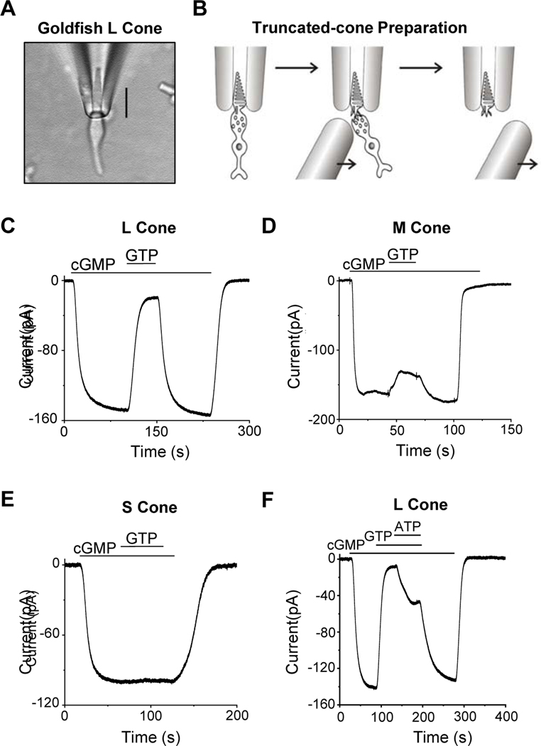
Figure 1. Truncated-cone recordings.
(A) Photomicrograph of a goldfish L cone with outer segment drawn into a suction-pipette electrode. (B) For truncated-cone recordings, a glass probe was used to separate the cone inner segment from the outer segment held in the pipette. After truncation, the intracellular compartment could be dialyzed through rapid solution changes. (C-E) Membrane current recorded from a truncated L (C), M (D) and S cone (E). The truncated cone was perfused continuously with a pseudo-intracellular solution, followed by addition of 500-μM cGMP which induced an inward current due to the opening of cyclic-nucleotide-gated (CNG) channels. Next, 15-μM GTP was added into the perfusion solution, leading to a large current reduction in L cones (C), a small current reduction in M cones (D), but no detectable reduction in S cones (E). In each cone subtype, the effects of cGMP and GTP could be completely washed out. (F) ATP effect on dark GTP-dependent PDE activity. Addition of 1-mM ATP inhibited GTP-induced current reduction in an L cone, presumably by quenching pigment activity via pigment phosphorylation.

