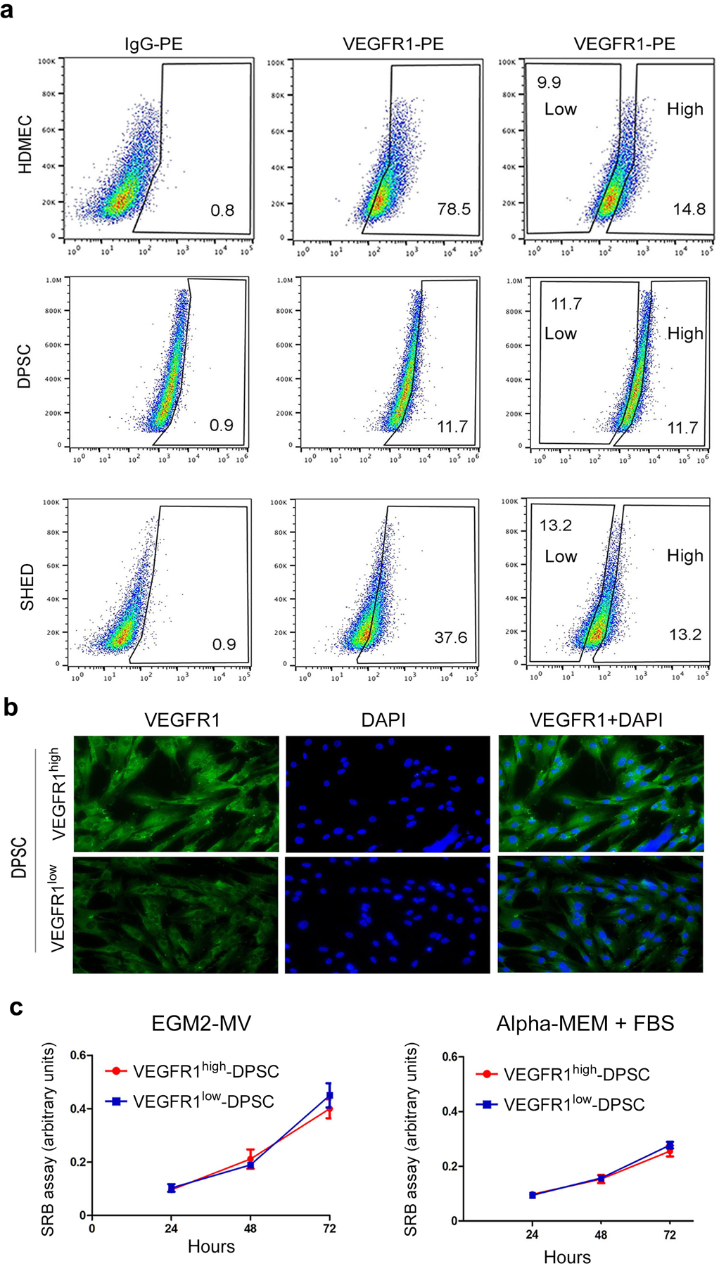
Fig. 2. VEGFR1 does not regulate proliferation of pulp stem cells.

(a) Flow sorting of HDMEC, SHED, and DPSC according to VEGFR1 expression levels (i.e. high and low), using isotype-matched IgG to set the gates. For DPSC and SHED, we sorted out equivalent percentages of VEGFR1high and VEGFR1low cells. (b) Fluorescence microscopy images of VEGFR1high and VEGFR1low cells. Green depicts VEGFR1 expression while blue depicts DAPI nuclear staining. bar=20 μm. (c) Line graph depicting cell proliferation over time when VEGFR1high and VEGFR1low cells, as determined by the SRB assay. Cells were cultured in vasculogenic differentiation medium (EGM2-MV + 50 ng/ml rhVEGF165) or alpha-MEM + 20% FBS for 24 to 72 hours. Data represents average +/− s.d. in 8 wells per condition.
