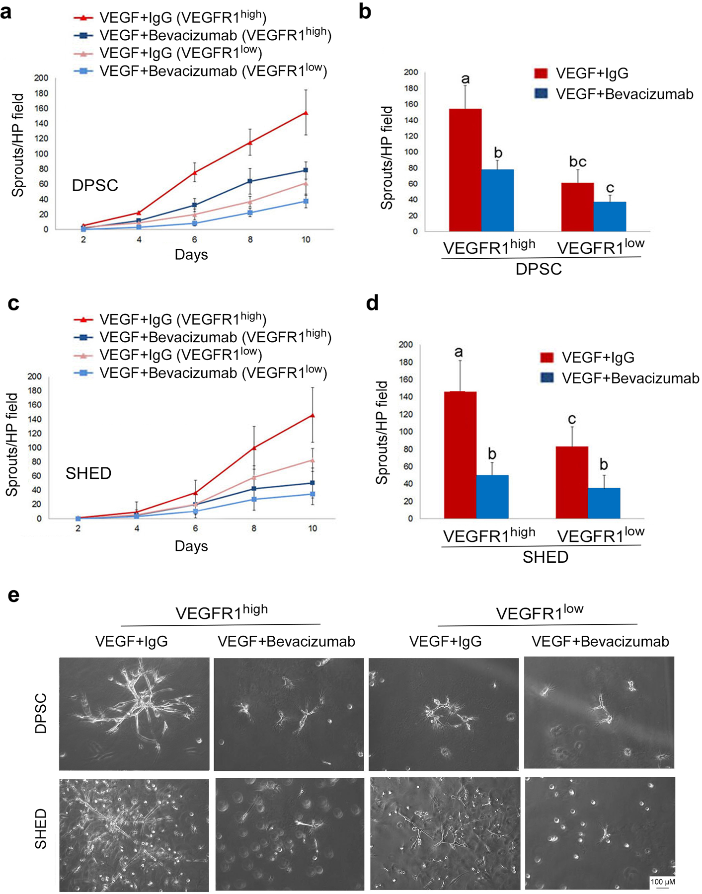
Fig. 4. VEGF blockade inhibits the vasculogenic potential of VEGFR1high cells in vitro.

(a,c) Line graphs depicting the number of sprouts per high power field generated by DPSC or SHED. (b,d) Bar graphs showing the number of capillary-like sprouts at the end of the experimental period (i.e. 10 days). VEGFR1high and VEGFR1low DPSC or SHED cells were cultured in wells pre-coated with growth factor reduced Matrigel and stimulated with vasculogenic differentiation medium in presence of 0 or 25 μg/ml bevacizumab (anti-VEGF antibody). Different low case letters indicate statistical significance at p<0.05. Number of capillary sprouts (average +/− s.d.) is representative of 12 random microscopic fields from triplicate wells per condition. (e) Representative photomicrographs of the capillary sprouts observed after 10 days under the experimental conditions described above (bar: 100 μm).
