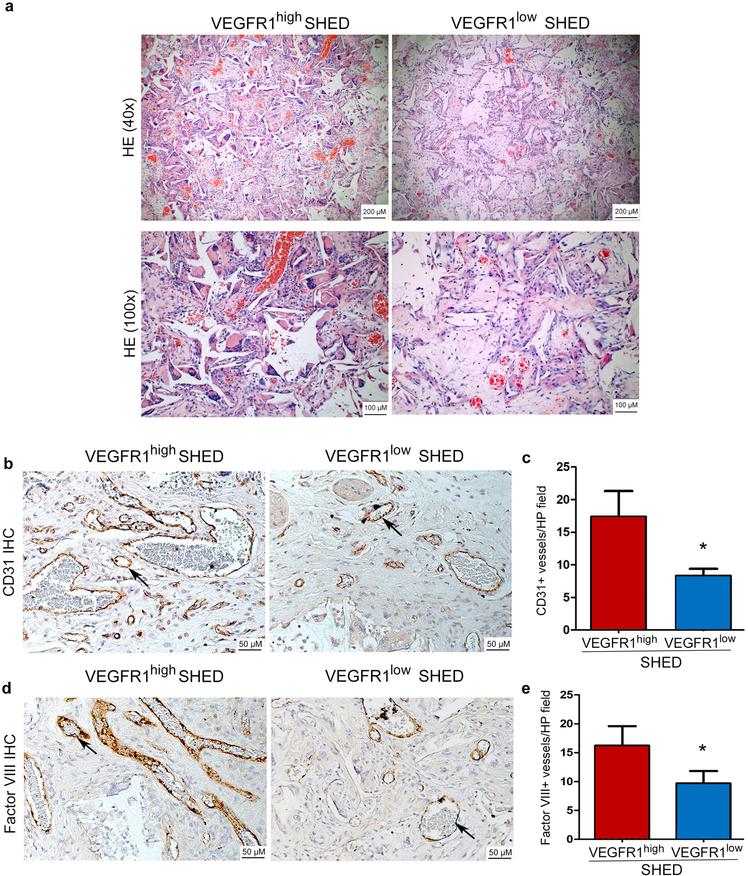
Fig. 5. VEGFR1high SHED cells are more vasculogenic than VEGFR1low SHED in vivo.

(a,c) Human VEGFR1high and VEGFR1low SHED were seeded in biodegradable scaffolds (n=6 per experimental condition) and transplanted into the subcutaneous space of immunodeficient mice. Four weeks after transplantation, the scaffolds were retrieved, fixed, and paraffin embedded. (a) representative images of sections stained with Hematoxylin and eosin at low and high magnification (bar: 100 μm/ 200 μm) and (b,d) Immunohistochemistry with anti-human CD-31 or anti-Factor VIII antibody to identify blood vessels (brown color). Representative vessels are highlighted with black arrows (bar: 50 μm). (c,e) Graphs depicting the number of CD31-positive or Factor VIII-positive blood vessels inside the scaffolds. Data represent analysis of 8 randomly selected microscopic fields from each scaffold (n=6) at 200x.
