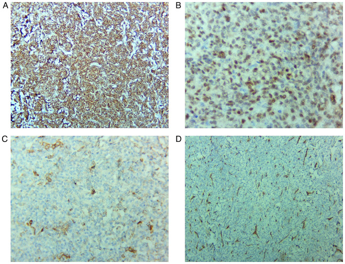
Figure 2.
Immunohistochemistry staining. (A) Intense, diffuse positivity for CD10; magnification, x100. (B) Tumor cells show nuclear positivity for ER, with variable intensity; magnification, x400. (C) Focal positivity for actin (clone HHF35); magnification, x200. (D) CD34 is negative in the tumor cells and positive in the blood vessels network; magnification, x100. ER, estrogen receptor. HHF35, anti-muscle actin antibody.

