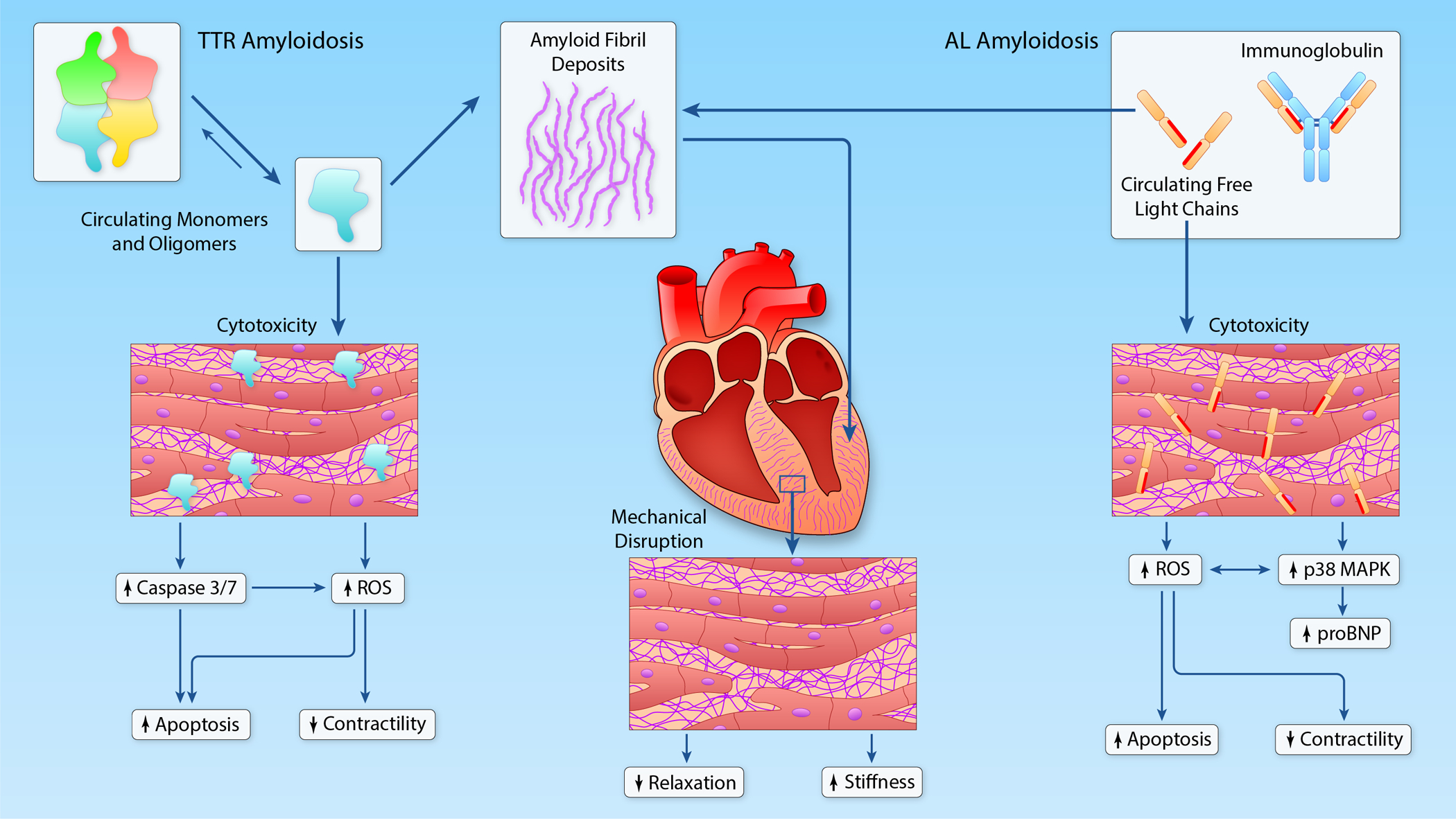
Figure 2:

Mechanisms of myocardial dysfunction in TTR and AL amyloidosis. In both TTR and AL, extracellular deposition of amyloid fibrils causes mechanical disruption of normal tissue architecture, leading to impaired relaxation and increased ventricular stiffness. In AL amyloidosis, circulating free light chains cause cytotoxicity by increasing oxidative stress and activation of the p38 MAPK signaling pathway. Activation of the p38 MAPK pathway also leads to release of NT-proBNP. In TTR amyloidosis, circulating TTR monomers and oligomers have also been proposed to cause direct cytotoxicity. (Illustration credit: Ben Smith).
