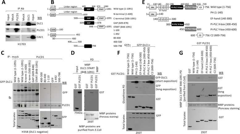
Fig. 1.
Mapping of DLC1 and PLCD1 interacting regions. A Co-immunoprecipitation of endogenous PLCD1, DLC1 and Caveolin-1 proteins in H1703 cells. The indicated antibodies for immunoprecipitation (IP) and Western blot (WB) were used. Input represents a fraction of the H1703 cell lysate before immunoprecipitation. B Schematic representation of the domain organization of DLC1 (top) and DLC1 constructs cloned as GFP-fusion proteins for immunoprecipitation in C. C PLCD1 Immunoprecipitation (IP) in cell lysates from H358 cells overexpressing control (GFP) or GFP-fusion plasmids spanning several regions in DLC1 followed by Western blot (WB) using the indicated antibodies. The expression of all GFP-fusion plasmids in total lysates is shown in the bottom panel. Asterisks (*) indicate the expression of the corresponding GFP-fusion proteins. D MBP pull-down (PD) in 293 T cells expressing GST control or GST PLCD1 using MBP control or MBP-DLC1 START 848–1091 purified proteins (from E.Coli), followed by GST Western blot (WB). The amount of MBP-purified proteins is shown as stained with Ponceau S (bottom panel). Input represents a fraction of the 293 T cell lysates before pull-down. E Schematic representation of the domain organization of PLCD1 (top) and PLCD1 constructs cloned as GST-fusion proteins to be used in F and G. F Glutathione pull-down (PD) in 293 T cells transfected with GST-PLCD1 plasmids encompassing several PLCD1 domains and GFP or GFP DLC1, followed by GFP and GST western blot. The expression of all GFP-fusion plasmids in total lysates is shown (bottom). G Pull-down (PD) using MBP DLC1 START 848–1091(purified from E. coli) and GST or GST-PLCD1 domains, followed by GST western blot. The amount of MBP- purified protein is shown as stained with Ponceau S. Expression of GST fusion proteins was checked by GST western blot in total lysates (bottom)

