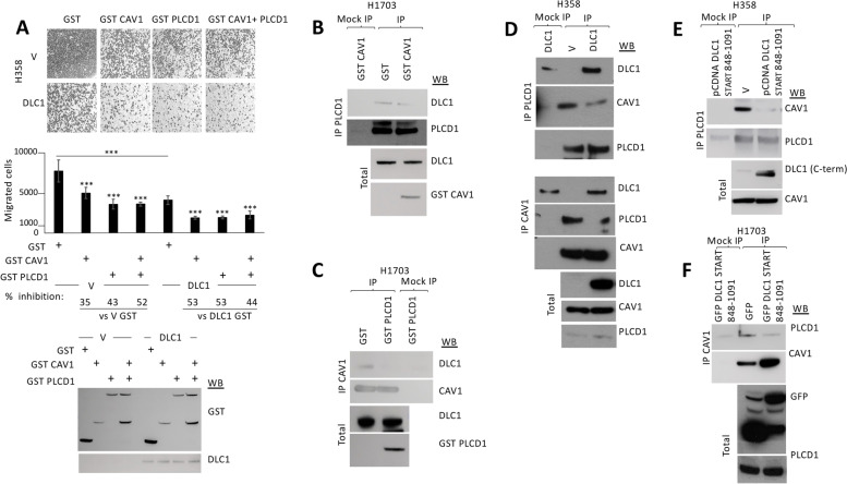
Fig. 2.
Caveolin-1 and PLCD1 cooperate with DLC1 to inhibit cell migration but their binding to DLC1 is mutually exclusive. A Migration in transwells of stable H358 control (V) or DLC1-overexpressing cells transiently transfected with GST control, GST Caveolin-1 (GST CAV1), GST PLCD1 or GST CAV1 + GST PLCD1. Migrated cells were fixed and photographed (top panel shows a representative image from each condition). The number of migrated cells were quantified using Image J (middle panel), and the percentage (%) of inhibition was calculated from a total of four random images taken from each experiment duplicate. Bars represent mean +/− SD. t-test was performed for statistical analysis (***:p < 0.005). The correct expression of GST proteins and DLC1 was checked by Western blot (WB). B PLCD1 Immunoprecipitation in H1703 cells transfected with GST control or GST Caveolin-1 (GST CAV1) plasmids, followed by Western blot (WB) using the indicated antibodies. C Caveolin-1 (CAV1) Immunoprecipitation (IP) in H1703 cells transfected with GST control or GST PLCD1 plasmids, followed by Western blot (WB) using the indicated antibodies. The correct expression of GST fusion proteins in B and C is shown from total lysates (bottom panels). D Immunoprecipitation (IP) using PLCD1 (top) or Caveolin-1 (CAV1, middle) antibodies in H358 cells transfected with empty vector (V) or pCDNA DLC1, followed by Western blot (WB) using the indicated antibodies. Correct expression of proteins in total lysates is shown in the bottom panel. E PLCD1 Immunoprecipitation (IP) in H358 cells transfected with empty vector (V) or a plasmid encoding the DLC1 START domain-containing region (DLC1 848–1091), followed by Western blot (WB) using the indicated antibodies. The correct expression of all proteins in total lysates is shown (bottom panel). F Caveolin-1 Immunoprecipitation (IP) in H1703 cells transfected with GFP or GFP-DLC1 START domain containing region (848–1091), followed by Western blot (WB) using the indicated antibodies. The correct expression of proteins in total lysates is shown (bottom panel)

