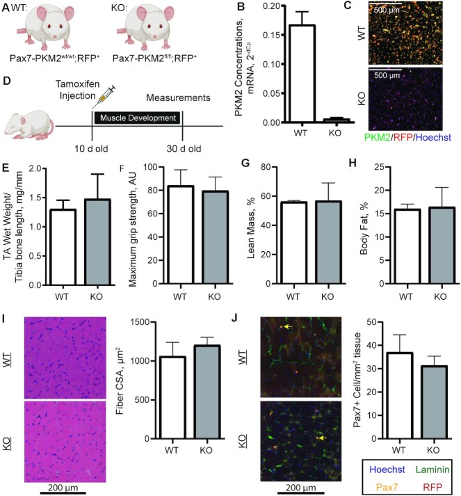
FIGURE 6.
Generation of an MPC-specific PKM2 KO mouse and measurements of muscle development. (A) Pax7-PKM2wt/wt; RFP+ (WT) and Pax7-PKM2fl/fl; RFP+ (KO) mice were generated. (B, C) Isolation of MPCs after in vivo tamoxifen injection confirmed successful PKM2 deletion (n = 2/genotype). (D) Ten-day-old pups were injected with tamoxifen and aged to 30 d of age for measurements (n = 7 WT and n = 4 KO). (E) TA wet weight normalized to tibia bone length, (F) maximum grip strength, (G) percentage lean mass, (H) percentage fat mass, (I) fiber CSA distribution, and (J) Pax7+ cell number were compared between WT (n = 7) and KO (n = 4) mice. Yellow arrows show Pax7+ cells. Values are mean ± SD. AU, arbitrary units; CSA, cross-sectional area; KO, knockout; MPC, muscle progenitor cell; Pax7, paired box 7; PKM2, pyruvate kinase M2; RFP, red fluorescent protein; TA, tibialis anterior; WT, wild-type.

