
Figure 4.
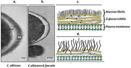
Visualizaton of Candida albicans cell walls grown in single-species (a) and dual-species biofilms (b). (Figures consisted of ≈100 cell images); bar, 5 nm. G + C, β-glucan and chitin; M, mannan. Drawings representing the possible structural changes are shown in c (for the cell wall of C. albicans in dual-species biofilm) and d (for the cell wall of C. albicans in single-species biofilm)