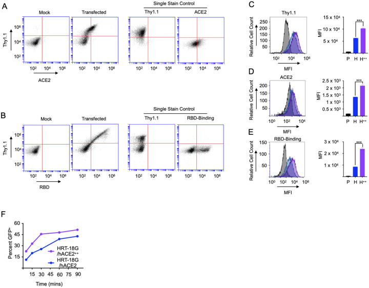
Figure 2. Increased ACE2 expression potentiates SARS-CoV-2 RBD binding and pseudoviral infectivity.
A, B. HRT-18G cells were transiently transfected with cDNA encoding hACE2 and Thy1.1 in a bi-cistronic cassette and two days later stained with (A) fluorescently labeled anti-Thy1.1 and ACE2 antibodies and analyzed by flow cytometry. Single antibody labeling controls are also depicted. B. Cells were stained with fluorescently labeled anti-Thy1.1 antibody and Alexa Fluor 647-labeled SARS-CoV-2 S-protein RBD analyzed by flow cytometry. Histograms of individual protein-labeled cells are also depicted. C. HRT-18G/hACE2 (blue trace) were fluorescently sorted based on Thy1.1 expression to generate the cell line HRT-18G/hACE2++ (purple trace). HRT-18G/hACE2 and HRT-18G/hACE2++, and parental HRT-18G (shaded histogram) cells were then incubated with (C) anti-Thy1.1, (D) anti-ACE2 antibodies, or (E) Alexa Fluor 647-labeled SARS-CoV-2 S-protein RBD and analyzed by flow cytometry. Representative histograms are shown as well as quantitative MFI measurements from three technical repeats. F. HRT-18G/hACE2 and HRT-18G/hACE2++ cells were infected with GFP-expressing SARS-CoV-2 pseudovirus for indicated times, washed to remove excess virus, and incubated in complete media for 16 hours and analyzed by flow cytometry for GFP expression. All data presented is representative of three independent experiments. Statistical significance is indicated as: *** P < 0.0001.

