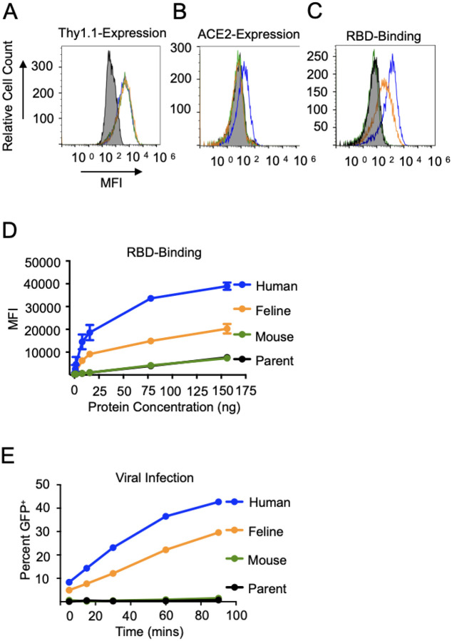
Figure 3. Equivalent expression of ACE2 orthologs and varied spike protein binding.
A. HRT-18G cells were stably transfected with IRES-Thy1.1 plasmids containing cDNAs of human (blue), feline (orange), and mouse (green) ACE2 and magnetically sorted until an equivalent level of reporter protein Thy1.1 was expressed on all three cell lines. B. To confirm the specificity of the ACE2 antibody for human ACE2, HRT-18G/hACE2, HRT-18G/fACE2, HRT-18G/mACE2 and HRT-18G (shaded histogram) cells were simultaneously incubated with fluorescently-labeled ACE2 antibody and analyzed by flow cytometry. C. The four cell lines incubated with 7.8 ng of Alexa Fluor 647-labeled SARS-CoV-2 S-protein RBD and analyzed for SARS-CoV-2 S-protein RBD/ACE2 binding affinity by flow cytometry. D. Same as in C except cells were incubated with different concentrations of Alexa Fluor 647-labeled RBD. The MFI of the population is reported on the y-axis. E. HRT-18G/hACE2, HRT-18G/fACE2, HRT-18G/mACE2 and HRT-18G cells were infected with GFP-expressing SARS-CoV-2 pseudovirus for specified times, washed to remove excess virus, and incubated for 16 hours, and GFP expressing cells quantified by flow cytometry. All data presented is representative of three independent experiments.

