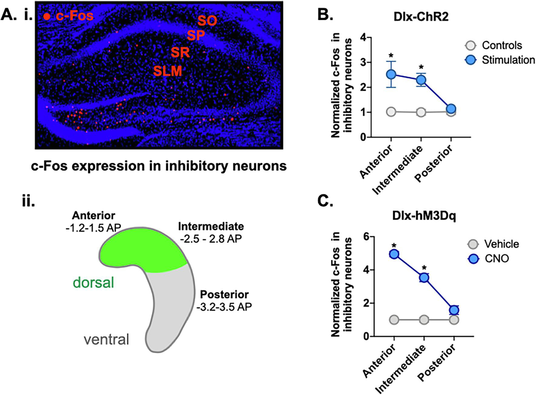
Figure 4. Widespread activation of inhibitory neurons in the dorsal hippocampus.

A. i. Anterior dHPC of representative CNO-treated Dlx-hM3Dq animal showing c-Fos expression (red) in inhibitory strata: stratum oriens (SO), stratum radiatum (SR), stratum lacunosum-moleculare (SLM). A. ii. Schematic of HPC showing location of virus expression (green) in dorsal HPC. Representative slices were taken for c-Fos analysis from anterior (−1.2 to −1.5 AP), intermediate (−2.5 to −2.8 AP) and posterior/ventral HPC (−3.2 to −3.5 AP). B. c-Fos expression in inhibitory neurons of anterior and intermediate HPC was increased in Dlx-ChR2 laser-stimulated animals (blue). There was no difference in c-Fos expression in posterior HPC. C. c-Fos expression in inhibitory neurons of anterior and intermediate hippocampus was increased in Dlx-hM3Dq CNO-treated animals (blue). There was no difference in c-Fos expression in posterior HPC. All data are expressed as mean +/− SEM.
