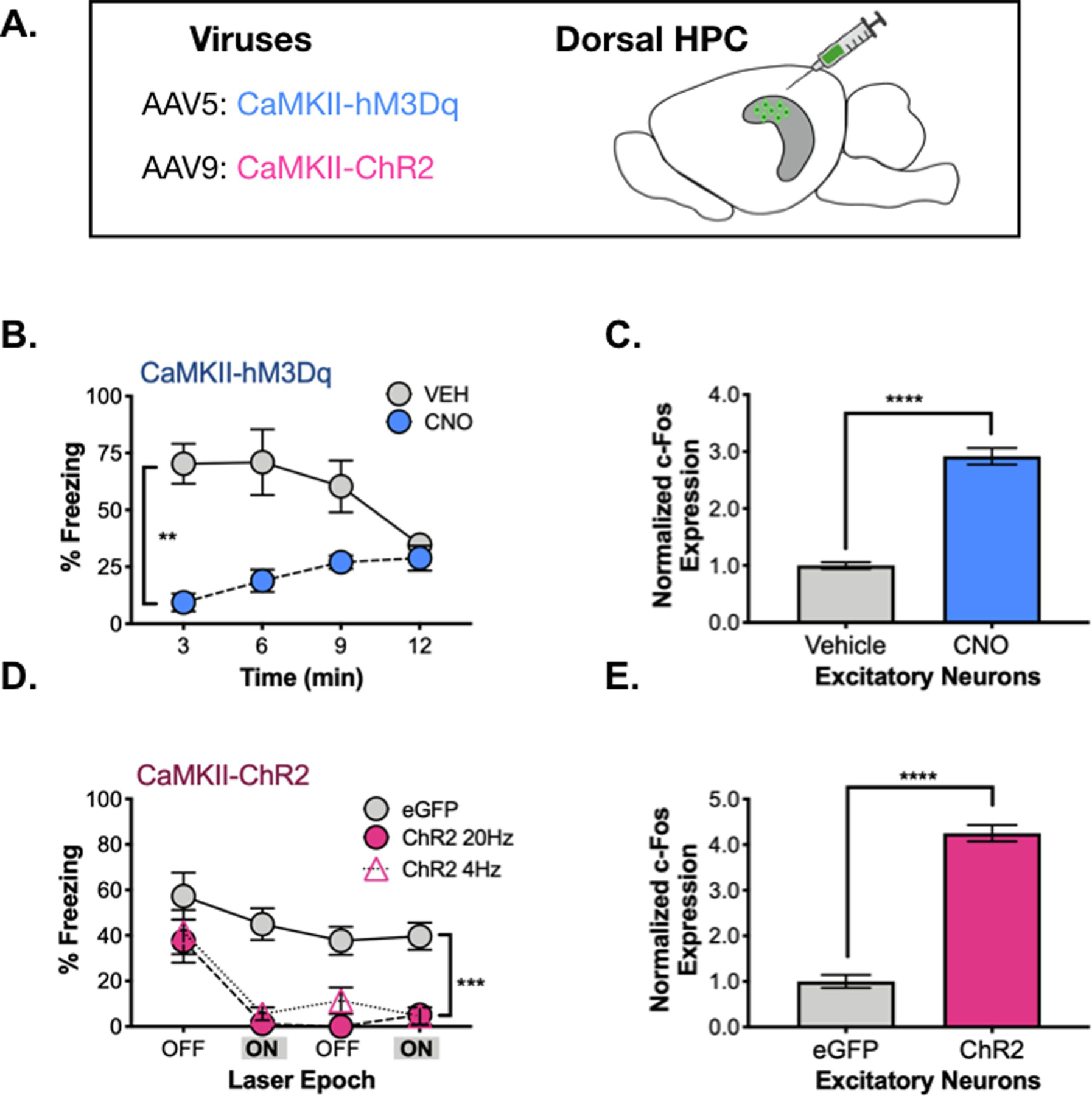
Figure 5. Prolonged vs. acute activation of excitatory neurons.

A. CaMKII-ChR2 or CaMKII-hM3Dq were infused into dHPC. B. During memory testing, hM3Dq CNO-treated animals (pink) froze less than their vehicle-treated counterparts (gray). C. c-Fos expression in excitatory neurons of dCA1 was elevated in hM3Dq CNO-treated animals (pink) compared to their vehicle-treated counterparts (gray). D. During memory testing, ChR2 (20Hz – pink circles, 4Hz – pink triangles) animals and eGFP animals (gray) did not differ during the first 3-minute laser OFF epoch. Following laser stimulation, 20 and 4Hz stimulated animals froze less than controls over the remainder of the testing period. E. c-Fos expression was elevated in excitatory neurons of dCA1 in ChR2 20Hz-stimulated animals (pink) compared to controls (gray). All data are represented as mean +/− SEM.
