. 2021 Sep 25;38(3):345–353. doi: 10.5511/plantbiotechnology.21.0727a

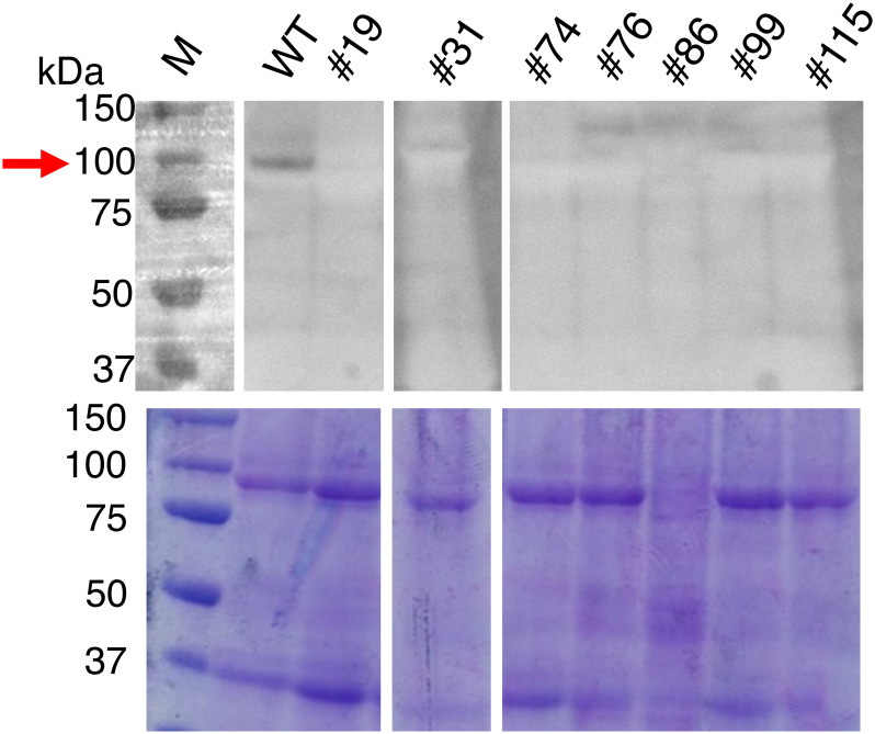
Figure 6. Detection of potato SBE3 in the tubers. Total protein was prepared from the tubers of the mutant lines and WT. Proteins were separated by SDS-PAGE (lower panel), and SBE3 was detected by western blot analysis using a rabbit antiserum raised against rice BE1 (upper panel). Gaps are introduced between the lanes of the gel. An arrow indicates the band corresponding to SBE3. Size markers are shown to the left. WT: wild-type, Numbers with the prefix #: mutant lines.

Figure 6. Detection of potato SBE3 in the tubers. Total protein was prepared from the tubers of the mutant lines and WT. Proteins were separated by SDS-PAGE (lower panel), and SBE3 was detected by western blot analysis using a rabbit antiserum raised against rice BE1 (upper panel). Gaps are introduced between the lanes of the gel. An arrow indicates the band corresponding to SBE3. Size markers are shown to the left. WT: wild-type, Numbers with the prefix #: mutant lines