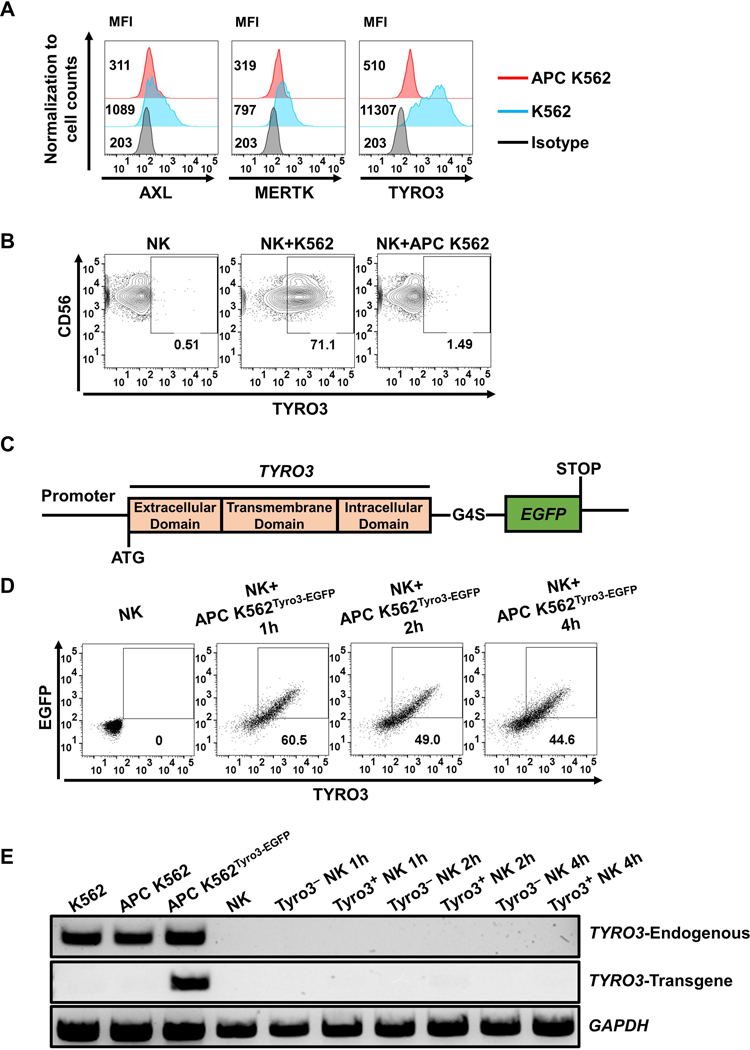
Figure 3. Tyro3-EGFP can be transferred from tumor cells to human NK cells via trogocytosis.
(A) Representative flow cytometry shows TYRO3, AXL and MERTK expression on parental K562 and APC K562 cells, confirming that the APC K562 cells barely express TYRO3. (B) NK cells did not express TYRO3 after co-cultured with APC K562 cells at an E/T ratio of 1:1 for 1 h. Each experiment was performed with 4 different donors. (C) The construct design of the Tyro3-EGFP fusion protein. G4S linker, Gly-Gly-Gly-Gly-Ser. (D) Representative flow cytometry plots showing co-culture of NK cells with APC K562 cells overexpressing TYRO3 with C-terminal fusion EGFP (APC K562Tyro3-EGFP). Each experiment was performed with NK cells from 3 different donors. (E) PCR analysis of TYRO3 expression in NK cells after co-cultured with APC K562Tyro3-EGFP cells. K562 cells were used as positive control for endogenous TYRO3 transcript and served as negative control for transgenic TYRO3 transcript. TYRO3− and TYRO3+ NK cells both lacked the expression of endogenous and transgenic TYRO3 transcript over the entire 4 h period of co-culture. GAPDH was used as control for quality of cDNA synthesis. Each experiment was performed with 3 different donors.

