Figure 7.
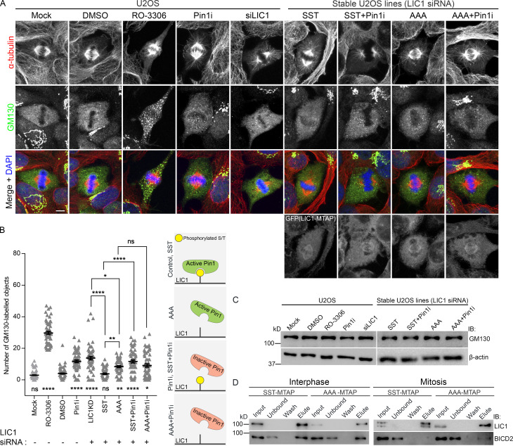
LIC1-CTD phosphorylation and Pin1 function regulate mitotic Golgi fragmentation. (A) Representative confocal images showing the localization of the Golgi protein GM130 in metaphase (after single thymidine block and release into MG132 for 2 h) under the indicated conditions. (B) Quantification of the number of GM130-positive punctae. n = 3 experiments, 20 cells per experiment. (C) Immunoblots showing the expression of GM130 under the indicated conditions. (D) Immunoblots from SBP-affinity precipitates of SST-MTAP or AAA-MTAP cell lysates at the indicated stages. IB, immunoblot. Scale bar = 10 µm. Error bars = mean ± SEM. *, P < 0.05; **, P < 0.01; ****, P < 0.0001 (Kruskal–Wallis test).

