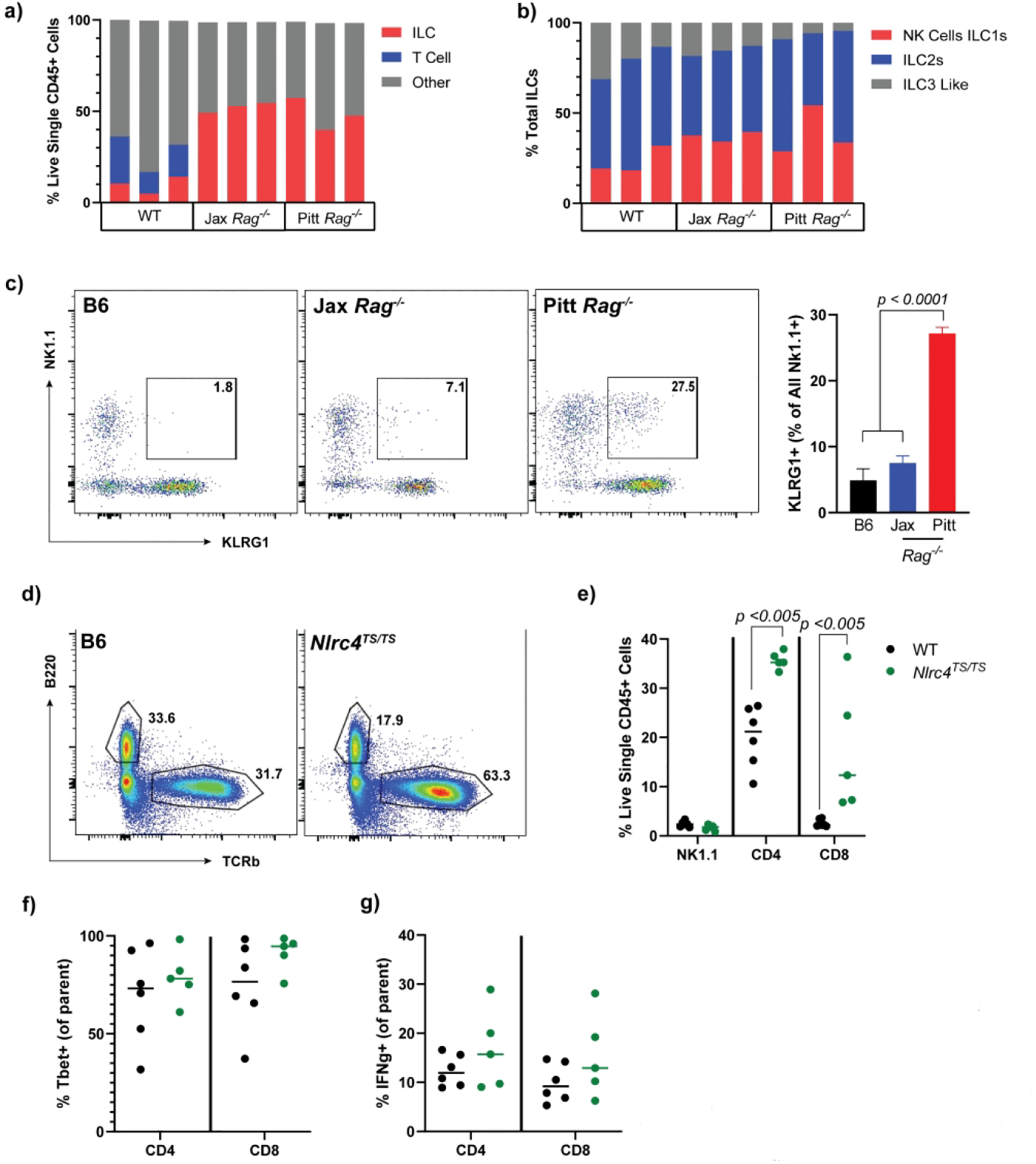
Figure 8: Elevated IL-18 alters lamina propria immune cell composition in Rag1−/− and NLRC4TS/TS mice.

Quantification of lamina propria lymphocytes by flow cytometry. Total live single CD45+ (a) and ILC (b) cell populations in Jackson WT vs Jackson and Pitt Rag1−/− mice. Representative flow plots and quantification showing KLRG1 NK1.1 double positive cells (c). T cell and NK cell composition (d and e) including representative flow plots and Tbet+ cells (f) in in house WT vs NLRC4TS/TS. Isolated ILCs were stimulated with Brefeldin, PMA and ionomycin for 2.5 hours and IFNg expression quantified (g). Panels a-c are representative on n=2 experiments with 3 mice per experimental group. Panels d-g show combined results from n=3 experiments with n=1–2 mice per group. Statistical significance is shown on each graph and was determined by unpaired t-test. Jax= Jackson Rag1−/− mice.
