
Fig. 1.
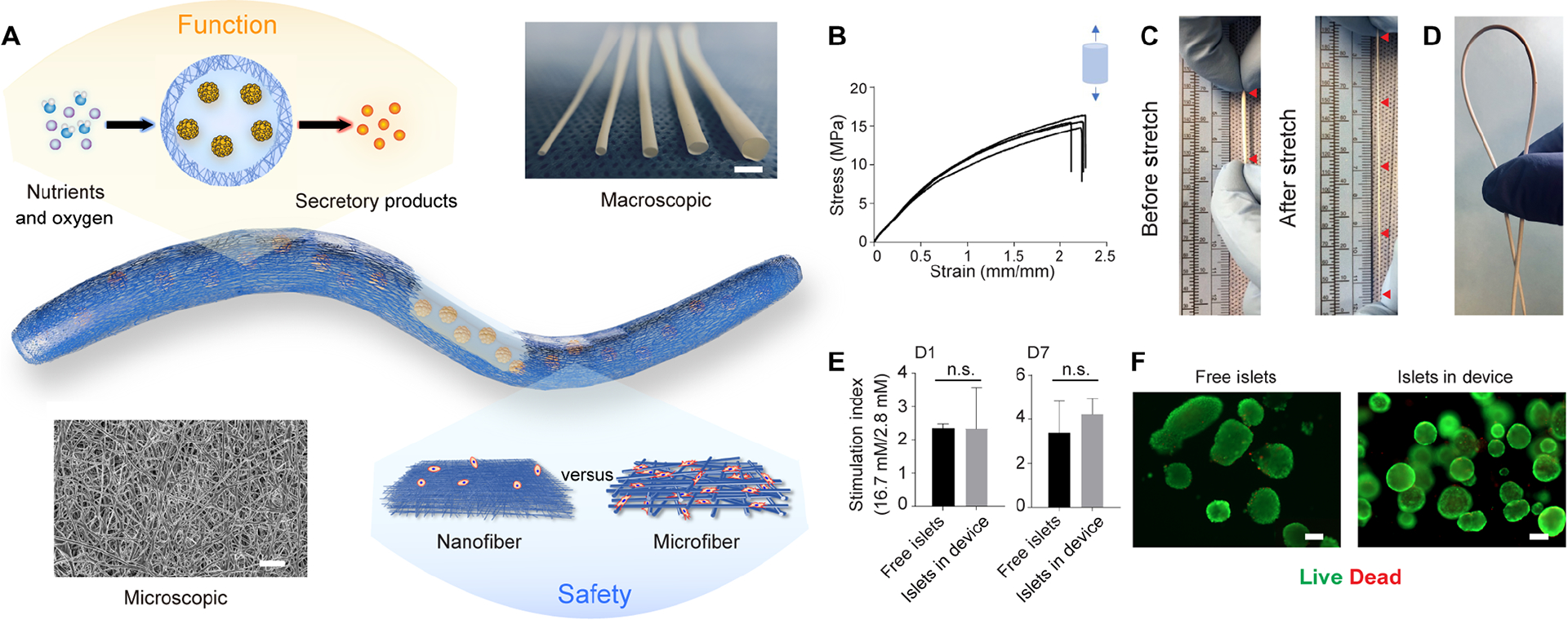
Design and characterization of the NICE device including mechanical properties, permeability and cell compatibility. (A) Schematics of the device showing the islet-laden hydrogel core surrounded by the nanofibrous skin that prevents cell penetration while allowing maximum mass transfer, along with a photo of nanofibrous tubes with different diameters (from left to right: 0.5 mm, 1 mm, 1.5 mm, 2 mm and 3 mm) and a SEM image of the nanofibers. (B) Tensile test (Stress-strain curves) of the nanofibrous tubes (n = 4). (C and D) Digital images showing the device being stretched more than three times (C) and bent without kink (D). (E) Stimulation index of islets (the ratio of insulin secretion in the buffers of high and low glucose concentrations) encapsulated in the device, compared to that of free-floating islets after 1-day and 7-days culture, mean ± SEM (n = 3). (F) Live (green) and dead (red) staining of free-floating islets and islets encapsulated in device after 1-day culture. The data was compared using the two-tailed Student’s t-test. The level of significance was labeled by n.s., denoting non-significant. Scale bars: 3 mm (A) macroscopic image, 100 μm (F) and 5 μm (A) microscopic image.