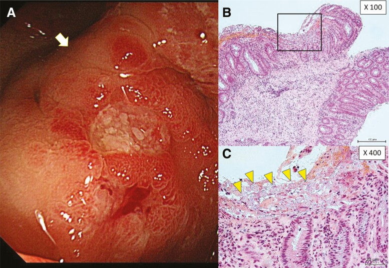
Figure 1.
Endoscopic and histopathologic findings from asymptomatic Entamoeba histolytica infections. A, Aphthae and slightly exudated lesions were observed in the cecum by endoscopy. Biopsy was performed at the edge of the aphthous lesion (white arrow). B and C, Trophozoite forms with phagocytosis of red blood cells [yellow arrowheads (C)] were detected on the membrane surface by hematoxylin and eosin staining. Trophozoites could not be identified in the submucosa.

