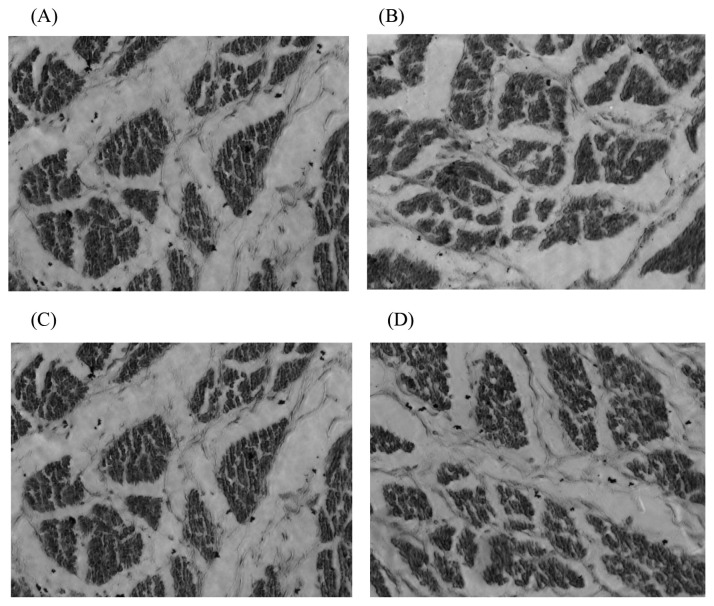
Figure 3.
The paraffin sections of breast muscle fiber in the 15th day embryo in 400× AMG EVOS microscope. (A) 100% energy group; (B) 80% energy group; (C) 70% energy group; (D) 50% energy group. Breast muscles were dehydrated in alcohol and finally embedded in paraffin wax. Transverse sections 10 mm thick were cut, stained with haematoxylin and eosin, and mounted in the usual way. Subsequently, 8 photomicrographs for fibre diameter measurements were taken using the AMG EVOS microscope’s particular morphometric function. In each muscle sample, more than fifty diameters were measured, and the densities were calculated. Compared with the 100% energy group, in general, the diameter and density of the embryonic breast muscle fibres in each energy-restricted group showed a decreasing trend.

