
Fig. 5.
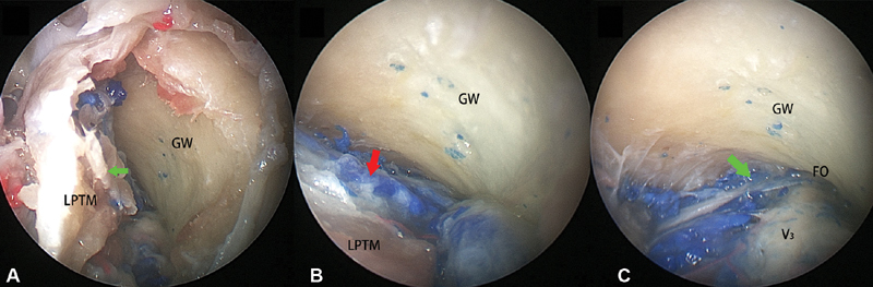
( A ) The superior head of the LPTM is elevated from the greater wing of sphenoid in a subperiosteal fashion (green arrow). ( B ) The pterygoid venous plexus (red arrow) located deep to the LPTM. ( C ) The communicated venous plexus (green arrow) connecting the pterygoid venous plexus and the cavernous sinus crossing the foramen ovale was present. LPTM, lateral pterygoid muscle.