Figure 1.
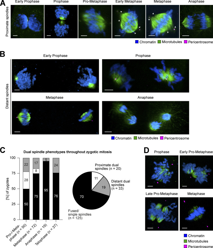
Dual spindle phenotypes in bovine zygotes. (A and B) IF of bovine zygotes fixed at 27.5 h after IVF at consecutive stages of mitosis. Maximum intensity projections orthogonal to the estimated spindle axis of confocal sections showing proximate (A) or distant (B) dual spindles. Shown are microtubules (α-tubulin, green), pericentrosomes (Cep192 or Nedd1, magenta), and chromatin (Hoechst, blue). Scale bars, 5 µm. White arrowheads indicate distinct pole clustering for proximate dual spindles. (C) Bar graph shows abundance (%) of dual spindle types at different mitotic stages. Pie chart summarizes abundance (%) of dual spindle types throughout mitosis. (D) IF staining of bovine zygotes as in A, but following a cold shock on ice for 3 min before fixation. Maximum intensity projections of confocal sections orthogonal to the estimated spindle axis showing that centrosomal microtubules have been depolymerized below the detection limit. Shown are microtubules (α-tubulin, green), pericentrosomes (Nedd1, magenta), and chromatin (Hoechst, blue). Scale bars, 5 µm.

