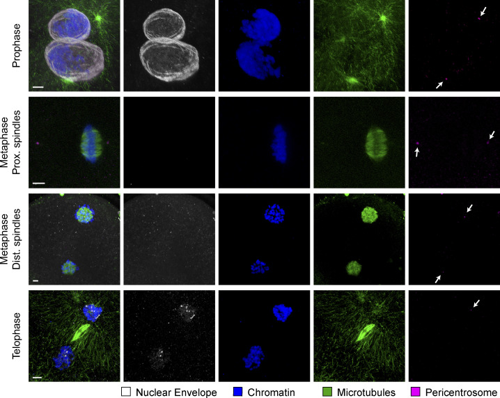
Figure S2.
Localization of nuclear lamina during first mitotic division in the bovine zygote. IF staining of bovine zygotes fixed at 27.5 h after IVF showing NE localization in consecutive stages of mitosis, from prophase to telophase and in proximate (Prox.) and distant (Dist.) dual spindles. Maximum intensity projections of confocal sections of the spindle volumes are shown for microtubules (α-tubulin, green), pericentrosomes (Nedd1, magenta), chromatin (Hoechst, blue), and NE (lamin B2, gray). Scale bars, 5 µm. Arrows indicate pericentrosome positions.

