Fig. 1.
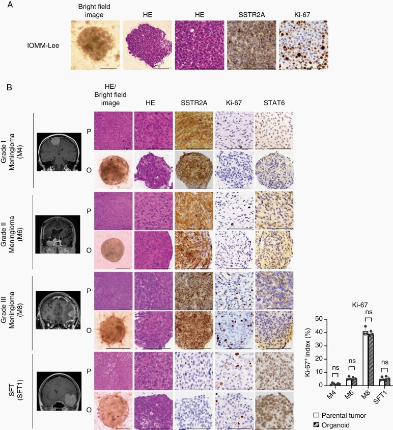
Established meningioma and SFT organoids recapitulate the histologic features of corresponding parental tumors. (A) Bright-field images, HE staining and IHC for anti-SSTR2A and anti-Ki-67 antibodies in the organoids (IOMM-Lee). Scale bars indicate 100 μm. (B) Magnetic resonance images, bright-field images, HE staining, and IHC using anti-SSTR2A, anti-Ki-67, and anti-STAT6 antibodies with Grades I (M4), II (M6), and III (M8) meningiomas and an SFT (SFT1) as parental tumors (P) and the organoids (O). Scale bars indicate 100 μm. Bar graph (right) indicating Ki-67 expression status. Ki-67 positive index was calculated and averaged in three fields. Average cell counts in a field were 130 (P) and 143 (O). The y-axis indicates Ki-67 positive index. Error bars indicate SD (ns: not significant).

