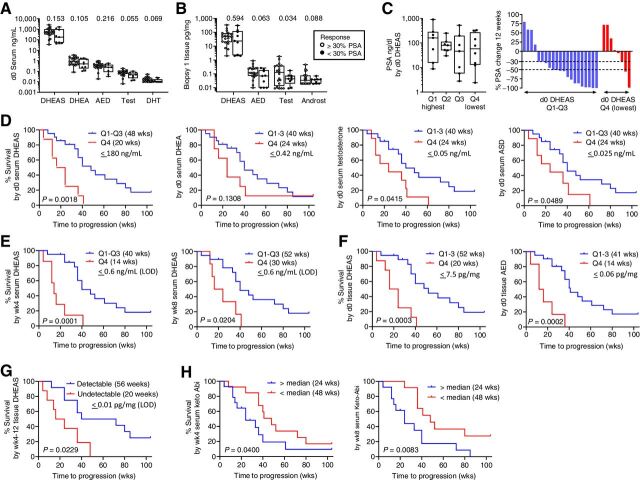
Figure 4.
Association of steroid and abiraterone levels with PSA decline and radiographic progression on AA plus prednisone. A and B, Comparison of steroid levels in serum (A) and metastatic tissue biopsies (B), based on achieving a 30% PSA decline at 12 weeks. C, Distribution of pretreatment PSA levels and waterfall plot showing percent change in PSA at 12 weeks by quartile (Q1-Q4) of pretreatment serum DHEAS levels. P values for the indicated comparison calculated via nonparametric Wilcoxon rank–sum test (Mann–Whitney test). D, Radiographic PFS (rPFS) as a function of baseline serum androgen levels comparing subjects in the lowest vs. highest 3 quartiles (Q4 vs. Q1–3). E, rPFS as a function of on-treatment serum DHEAS levels at week 4 (wk4) and week 8 (wk8) comparing subjects in the lowest vs. highest 3 quartiles (Q4 vs. Q1–3). F, rPFS as a function of pretreatment tissue DHEAS and AED levels comparing subjects in the lowest vs. highest 3 quartiles (Q4 vs. Q1–3). In each case, the cut-off value reflects the highest number of the bottom one-fourth of the values. The quartiles were separately assessed in the pre- and on-treatment populations. G, rPFS comparing subjects with detectable vs. undetectable levels of DHEAS in tissue biopsies taken at 4 and 12 weeks of therapy. H, rPFS as a function of serum 3-keto-5α-abiraterone levels (keto-Abi) at wk4 and wk8 comparing subjects above vs. below the median. PFS was estimated using Kaplan–Meier methods and compared using the Gehan–Wilcoxan test. Androst, androsterone; Q, quartile.

