Figure 1.
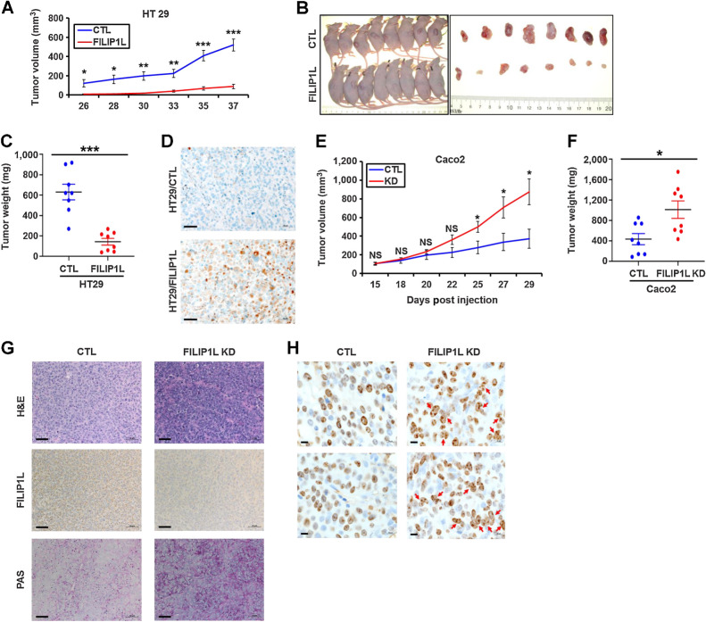
FILIP1L levels affect colon xenograft tumor growth, and its knockdown induces mucin secretion as well as multinucleation in colon xenograft tumors. A, HT29 (1.5 × 10⁶) clones of either control or FILIP1L+ derivatives were subcutaneously injected into the nude mice (8 mice per cell line). Tumor growth was measured, every 2 to 3 days for a total of 37 days. The y-axis represents tumor volume that was calculated by the formula: (length × width × height × 0.52). B and C, Pictures of mice and HT29 xenograft tumors at the time of sacrifice (B) as well as tumor weights (C) are shown. D, HT29 xenograft tumors from either control or FILIP1L+ derivatives were fixed and IHC stained for FILIP1L. E–H, Caco2 (5 × 10⁶) clones of either control or FILIP1L-knockdown derivatives were subcutaneously injected into the nude mice (8 mice per cell line). E and F, Tumor growth (E) was measured every 2 to 3 days for a total of 29 days, as described in A, and tumor weights at the time of sacrifice (F) were measured. G, Caco2 xenograft tumors from either control or FILIP1L-knockdown derivatives were fixed and stained with H&E and PAS. They were also IHC stained for FILIP1L. Scale bar, 50 μm. H, Enlarged images of Ki67-stained Caco2 xenograft tumors from either control or FILIP1L-knockdown derivatives are shown. Arrows, clumpy multinucleated cells. Scale bar, 10 μm. *, P < 0.05; **, P < 0.01; ***, P < 0.001.

