. 2021 Aug 20;81(21):5523–5539. doi: 10.1158/0008-5472.CAN-21-0897

Figure 5.

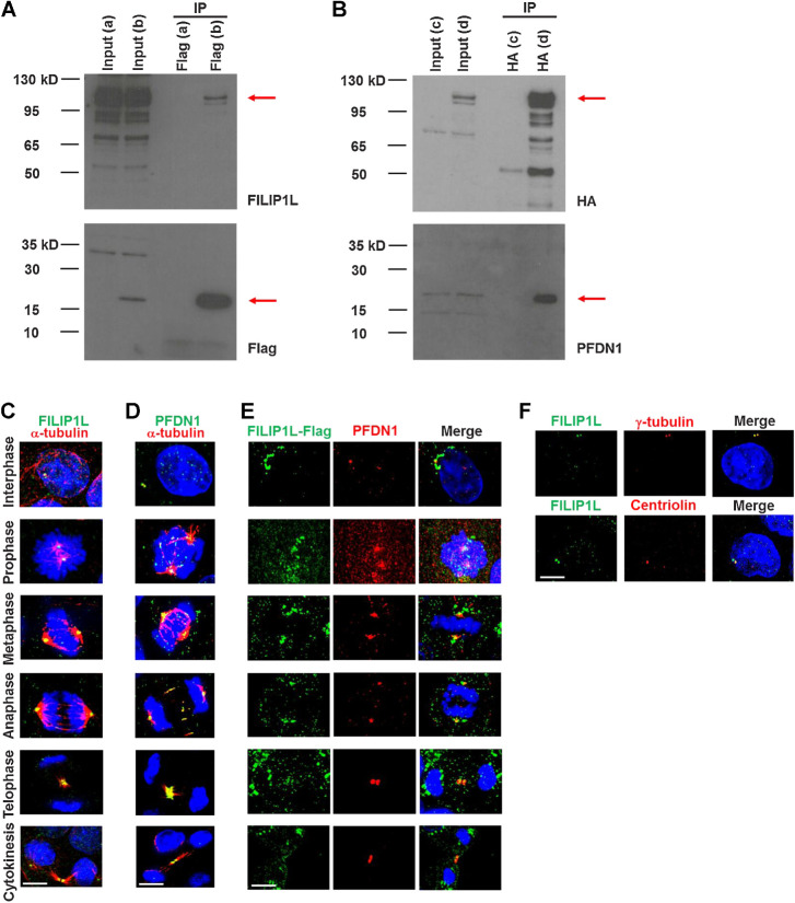
Figure 5. FILIP1L binds to and colocalizes with its binding partner PFDN1 at centrosomes in each phase of mitosis. HEK293T cells were transfected with either FILIP1L-HA and control-Flag vector (a), FILIP1L-HA and Flag-PFDN1 (b, d), or control-HA vector and Flag-PFDN1 (c). Twenty-four hours later, cell lysates were immunoprecipitated using Flag antibody-agarose, followed by immunoblotting for FILIP1L and Flag tag (A) or using HA antibody-agarose followed by immunoblotting for HA tag and PFDN1 (B). Input control (4 μg lysates) was also immunoblotted. C–F, MDCK.2 cells were immunofluorescently stained for FILIP1L (C) or PFDN1 (green; D) and α-tubulin (red). Nuclei were counterstained with DAPI (blue). Cell phase was determined by α-tubulin and DNA stain. A merged image is shown. E, MDCK.2 cells were transfected with a FILIP1L-Flag construct and stained for Flag tag (green) and PFDN1 (red) at 24 hours after transfection. F, MDCK.2 cells were stained for FILIP1L (green) and γ-tubulin or centriolin (red). Scale bar, 5 μm.

FILIP1L binds to and colocalizes with its binding partner PFDN1 at centrosomes in each phase of mitosis. HEK293T cells were transfected with either FILIP1L-HA and control-Flag vector (a), FILIP1L-HA and Flag-PFDN1 (b, d), or control-HA vector and Flag-PFDN1 (c). Twenty-four hours later, cell lysates were immunoprecipitated using Flag antibody-agarose, followed by immunoblotting for FILIP1L and Flag tag (A) or using HA antibody-agarose followed by immunoblotting for HA tag and PFDN1 (B). Input control (4 μg lysates) was also immunoblotted. C–F, MDCK.2 cells were immunofluorescently stained for FILIP1L (C) or PFDN1 (green; D) and α-tubulin (red). Nuclei were counterstained with DAPI (blue). Cell phase was determined by α-tubulin and DNA stain. A merged image is shown. E, MDCK.2 cells were transfected with a FILIP1L-Flag construct and stained for Flag tag (green) and PFDN1 (red) at 24 hours after transfection. F, MDCK.2 cells were stained for FILIP1L (green) and γ-tubulin or centriolin (red). Scale bar, 5 μm.