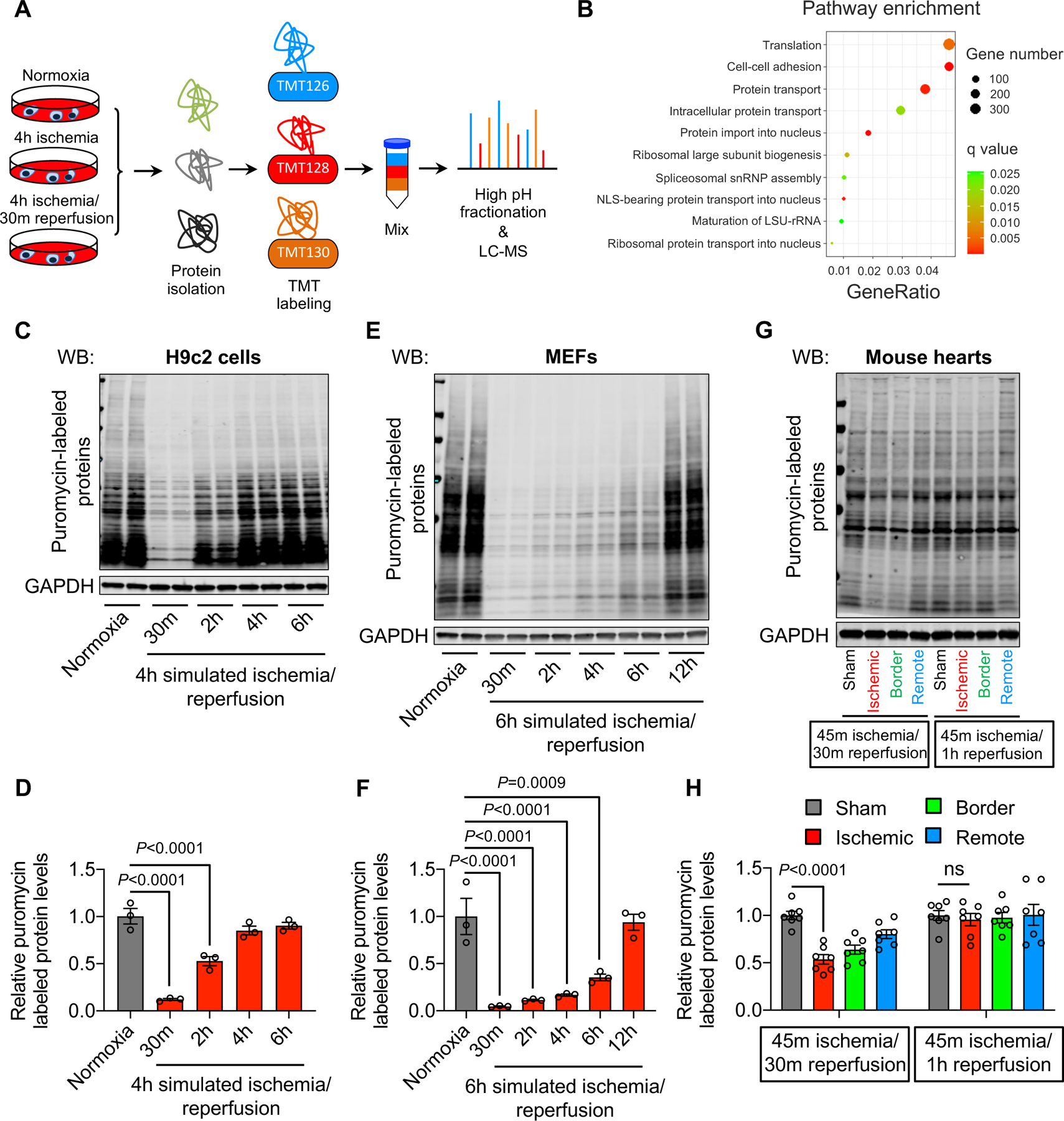
Figure 1. Ischemia/reperfusion is associated with a decrease in protein synthesis.

A. H9c2 cardiac myocytes were subjected to simulated ischemia (sI) for 4 hours or simulated ischemia for 4 hours, followed by reperfusion for 30 minutes (sI/R). Total proteins were isolated for tandem mass tag (TMT) labeling and mass spectrometry (MS).
B. Gene Ontology (GO) pathway analysis showed an enrichment of protein translation.
C. sI/R decreased protein synthesis. H9c2 cells were used for normoxia or sI/R. Puromycin was added 15 minutes before cell harvest. Total proteins were extracted to detect puromycin incorporation as a measure of protein synthesis.
D. Quantification of (C). n=3.
E. sI/R in mouse embryonic fibroblasts (MEFs) reduced protein synthesis. MEFs were subjected to either normoxia or sI/R. Puromycin was added 15 minutes before cell collection. Puromycin incorporation was detected by Western blotting.
F. Quantification of (E). n=3.
G. Ischemia/reperfusion (I/R) in vivo decreased protein synthesis in mouse hearts. Wild-type mice were subjected to cardiac ischemia for 45 minutes, followed by reperfusion for either 30 minutes or 1 hour. Puromycin was injected 30 minutes before heart collection. Cardiac tissues from different regions (ischemic, red; border, green; remote, blue) were separated and used for immunoblotting.
H. Quantification of (G). n=4.
One-way ANOVA was conducted, followed by Dunnett’s multiple comparisons test (D, F, H). Data are represented as mean±SEM. WB, Western blot and ns, not significant.
