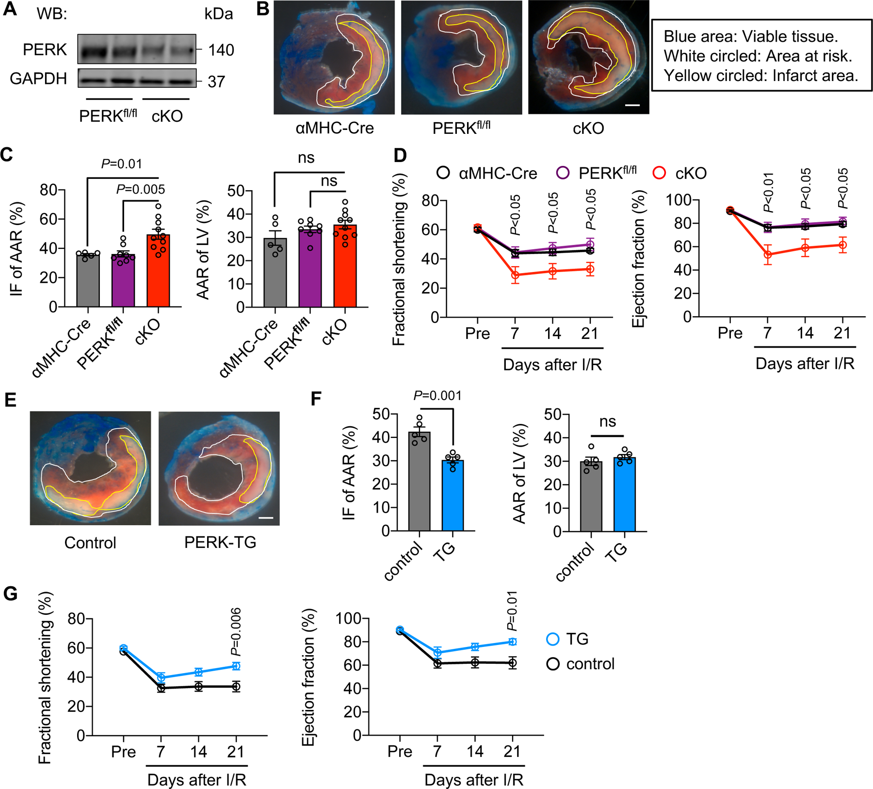
Figure 3. PERK protects the heart from reperfusion injury in vivo.

A. Validation of PERK knockout efficiency. Adult cardiomyocytes were isolated from PERKfl/fl and PERK cardiac-specific conditional knockout (cKO) mice, respectively, and used for immunoblotting.
B. PERK cKO mice and littermate controls were subjected to cardiac ischemia for 45 minutes, followed by reperfusion for 24 hours. The heart was then collected for triphenyltetrazolium chloride (TTC) staining to visualize viable tissue, area at risk, and infarct region. Different areas are circled. Scale bar: 1 mm.
C. PERK cKO led to an increase of infarct area/area at risk (IF/AAR). No difference in area at risk/left ventricle (AAR/LV) was found, suggesting that I/R surgeries were similarly conducted. n=5 to 10.
D. PERK cKO caused more severe reduction of cardiac function, as shown by decreases in fractional shortening and ejection fraction. n=5 to 7.
E. PERK transgenic (TG) mice and littermate controls were used for cardiac I/R. Dimer was administrated at the time of reperfusion. TTC staining was conducted. Control mice included αMHC-Cre only, CAG-FKBP-PERK only, and wild-type animals. Scale bar: 1 mm.
F. PERK activation conferred cardioprotection against I/R, as shown by a decrease of infarct size. Surgery was similarly conducted. n=5.
G. Cardiac function was significantly improved by PERK activation in the heart. n=6 to 9.
One-way ANOVA was conducted, followed by Tukey’s multiple comparisons test (C). Two-way ANOVA was conducted, followed by Tukey’s multiple comparisons test (D) and Sidak’s multiple comparisons test (G). Unpaired Student’s t test was conducted (F). Data are represented as mean±SEM. WB, Western blot and ns, not significant.
