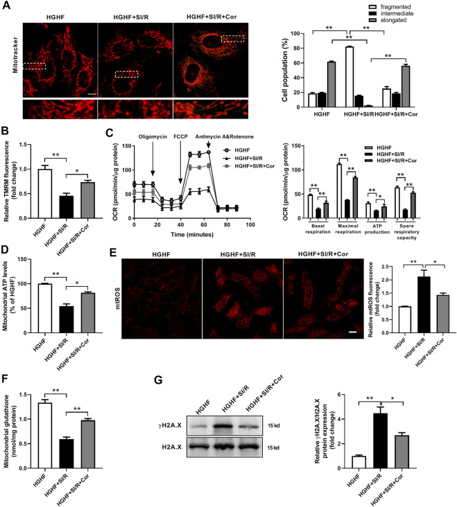
FIGURE 3.
Cordycepin enhanced Mfn2-medicated mitochondrial fusion and improved mitochondrial function in HG/HF cultured SI/R cardiomyocytes. (A) Representative images and quantitative analysis of mitochondrial morphology in cardiomyocytes under different treatments. Fifty to 60 cells per sample were counted. Scale bar, 10 μm. (B) Quantitative analysis of relative TMRM fluorescence that represented mitochondrial membrane potentials in cardiomyocytes under different treatments. (C) Measurement of Oxygen Consumption Rate (OCR) in H9c2 cardiomyocytes in different groups. n = 5 wells. (D) Measurement of the intracellular ATP levels in different groups. (E) Representative images and quantitative analysis of mtROS in cardiomyocytes under different treatments. Scale bar, 20 μm. (F) Mitochondrial glutathione content in mitochondria isolated from cardiomyocytes under different treatments. (G) Western blotting and quantitative analysis of γH2A.X and H2A.X in cardiomyocytes. n = 3 wells. HG/HF, high-glucose/high-fat; SI/R, simulated ischemia/reperfusion; Cor, cordycepin; TMRM, tetramethylrhodamine methyl ester. *p < 0.05, **p < 0.01. n = 6 wells.

