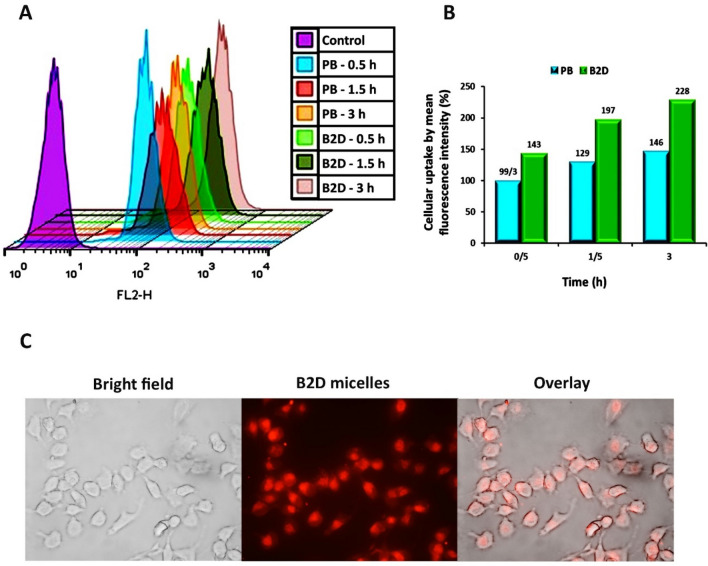
Figure 5.
(A) Results of intracellular uptake of rhodamine B-labeled blank βCD-g-PMA-co-PLGA micelles (PB: 20 µg/mL) and rhodamine B-labeled co-drug loaded βCD-g-PMA-co-PLGA micelles (B2D: 2 µg/mL) by MDA-MB-231 cells in different time intervals: 0.5, 1.5 and 3 h, using flowcytometry; (B) Diagram of mean fluorescence intensity (%) of rhodamine B-labeled PB and B2D that were uptaken by MDA-MB-231 cells in different time intervals: 0.5, 1.5 and 3 h, using flowcytometry; (the differences between treatments was statistically significant, p < 0.001); (C) Fluorescence microscopic images of rhodamine B-labeled B2D (2 µg/mL) internalization to MDA-MB-231 cells only at 1 h, prepared by research fluorescence microscope. (Abbreviations: PB: Rhodamine B-labeled blank βCD-g-PMA-co-PLGA micelles, B2D: Rhodamine B-labeled co-drug loaded βCD-g-PMA-co-PLGA micelles).

