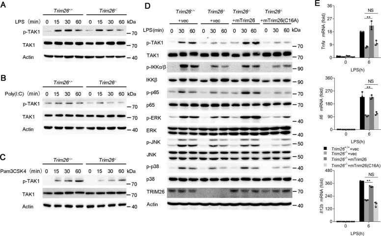
Fig. 5. TRIM26 enhances phosphorylation and complex assembly of TAK1.
A–C The Trim26+/+ and Trim26–/– peritoneal macrophages were stimulated with LPS (200 ng/ml), Poly(I:C) (20 μg/ml), or Pam3CSK4 (1 μg/ml) for the indicated times, and immunoblotting analysis were performed using anti-p-TAK1 or anti-TAK1 Abs. D Immunoblotting analysis of phosphorylated (p) and total TAK1, IKKα/β, p65, ERK, JNK, and p38 in Trim26+/+ and Trim26–/– peritoneal macrophages. Trim26–/– peritoneal macrophages reconstituted with vectors for mTrim26 or mTrim26(C16A) stimulated with LPS (200 ng/ml) for the indicated times. E qPCR analysis of Tnfα, Il6, and Il12b mRNA expression in Trim26+/+and Trim26–/– peritoneal macrophages. Trim26–/– peritoneal macrophages reconstituted with vectors for mouse mTrim26 or mTrim26(C16A) stimulated with LPS (200 ng/ml) for 6 h. Data are shown as mean ± SD of triplicates from one representative experiment in E. *p < 0.05, **p < 0.01 (one-way analysis of variance, ANOVA). Similar results were obtained in three independent experiments.

