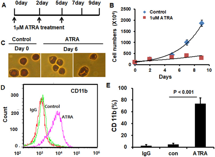
Figure 3.
HL-60 cells differentiated to neutrophil-like cells by all–trans-retinoic acid (ATRA). (A,B) Growth curve. HL60 cells (5 × 10⁵/mL) were seeded into a T-75 flask and treated with 0 or 1 μM ATRA once every 2 to 3 days for 5 days. Cells not stained by trypan blue were counted for 9 days. (C) Nuclear morphology of HL60 cells. HL60 cells were treated with 0 or 1 μM ATRA for 6 days and stained with Wright-Giemsa. At day 0, the nuclear morphology of dHL60 cells was ovate (left) but showed a dent (center) or lobular shape at 6 days (right) (magnification, ×400). (D,E) FACS analysis of CD11b expression in dHL60 cells. HL60 cells were differentiated by 0 or 1 μM ATRA for 6 days and stained with an anti-CD11b-FITC antibody. IgG was used as the negative control. Expression of CD11b (left). Percentage of total cells expressing CD11b (right). Independent sample t-test was used to find the difference.

