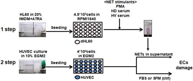
Figure 5.
Schematic diagram of the experimental procedure. HL-60 cells were seeded into T75 flasks at 5 × 10⁵ cells/mL and cultured for 6 days in 20% IMDM with 1 nM ATRA. Differentiated HL-60 cells were seeded into 24-well plates at 4.5 × 10⁵ per well and cultured in RPMI1640 with 1% BSA. PMA, HV serum and HD serum were applied to dHL-60 cells for 17 h and nuclease was added for 1 h. Cell free–NETs in supernatants were transferred to a new e-tube containing EDTA. HUVEC cells were cultured in 10% EGM2 and 4 × 10⁴ were seeded into 24-well plates. The day before the experiment, the culture medium was replaced with serum-free medium (SFM), 10% FBS, or 10–20% of NET supernatant. Cells were incubated for 20 h and used for analysis.

