Figure 6.
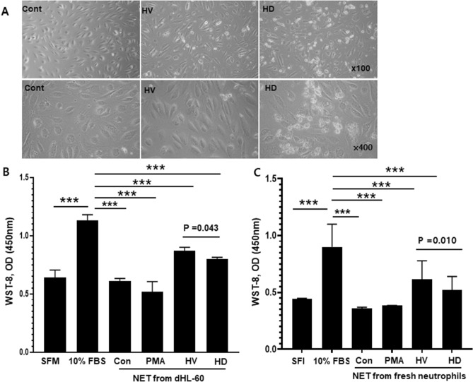
Damage to endothelial cells by NETs. To assess HUVEC damage, control (0 nM PMA), 400 nM PMA, HV serum, and HD serum were applied to dHL-60 cells for 17 h. Supernatants (10%) were added to HUVECs and incubated for 20 h. Serum-free medium (SFM) and 10% FBS were used as controls. (A) Morphology of HUVECs. In HV and HD NETs, cells became elongated compared to the control, and many dead floating cells were observed (magnification, upper panels, ×100; lower panels, ×400). (B) Effect of NETs from dHL-60 cells on HUVEC viability was tested by WST-8 assay. HUVEC viability was higher in HV serum-induced NETs than in uremic serum-induced NETs and SFM, and lower than those in 10% FBS. (C) Repeat experiments with isolated neutrophils from blood donor. Results are means ± SEM, Student’s t-test, ***p < 0.001.

