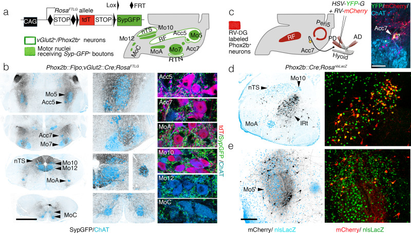
Fig. 1. Premotor status of reticular formation Phox2b+interneurons.
a RosaFTLG allele used for intersectional transgenic labeling of boutons from vGlut2/Phox2b interneurons (left) and schematic of the results (right). b Coronal sections through the hindbrain of a Phox2b::Flpo; vGlut2::Cre;RosaFTLG mouse at P4, showing synaptic boutons (black) from vGlut2/Phox2b interneurons in relation to motor nuclei (ChAT+, blue) at low (left), and higher (middle) magnifications, and close-ups of boutons (green) on motoneurons (right), which are either Phox2b+ (purple) or Phox2b− (blue). c (left) Strategy for mono-synaptically restricted transsynaptic labeling of premotor neurons from the posterior digastric muscle (PD) in a Phox2b::Cre;RosanlsLacZ mouse, with G-deleted rabies virus (RV) encoding mCherry and complemented by a G-encoding helper HSV virus (HSV-YFP-G), and summary of the results. (right panel) The only seed neurons are Acc7 motoneurons, double-labeled by the HSV-G and RV-mCherry viruses. d, e Coronal sections through the hindbrain at P8 showing labeled premotor neurons (black on the left panels) in the IRt (d) and Peri5 (e), which for the most part (72.7% ± 3.5 SEM, n = 4 animals) express Phox2b (right panels). AD anterior digastric, IRt intermediate reticular formation, nTS nucleus of the solitary tract, PD posterior digastric, Peri5 peritrigeminal area, RF reticular formation, RTN retrotrapezoid nucleus. Scale bars, b 1 mm for the left column, c 250 μm, d, e 500 μm.

