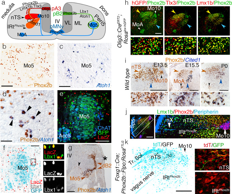
Fig. 2. Ontogenetic definition of IRtPhox2b and Peri5Atoh1.
a Two schematic hemisections of the embryonic medulla (left) or pons (right), showing the origin of branchiomotor nuclei (Mo5, MoA, and Mo10), Peri5Phox2b and IRtPhox2b in progenitor (p) domains of the ventricular layer (VL), their settling sites in the mantle layer (ML), and their transcriptional codes. b–d Coronal sections through the pons at E18.5, showing Peri5Phox2b (b) or Peri5Atoh1 (c, d) labeled with the indicated antibody or probe. Peri5Atoh1 cells co-express Phox2b and Atoh1 (arrowheads in d). e Coronal sections through Mo5 in a Phox2b::Flpo;Atoh1::Cre;Fela mouse at P0, showing the doubly recombined (nlsLacZ+) cells of Peri5Atoh1 (red). f Coronal section through Mo5 in a Phox2b::Flpo;Atoh1::Cre;Fela mouse, where Phox2b+ motoneurons are GFP+ (cyan) and Phox2b+/Atoh1+ neurons are nlsLacZ+ (red), counterstained for Lbx1 (gray at low magnification, green in the close-ups). g Coronal section through the pons at E11.5 showing the migrating Phox2b+ Mo5 and dB2 precursors (black and brown arrowheads, respectively) and, at their meeting point, Peri5Atoh1 cells that have switched on Atoh1. Asterisk: lateral recess of the IVth ventricle (IV). h Coronal sections through nTS (yellow arrowhead) and IRtPhox2b (blue arrowhead) at E18.5, at low magnification (upper) or at high magnification for the IRt (lower), stained with the indicated antibodies. A history of Olig3 expression is revealed by recombination of the histone-GFP (hGFP) reporter in the Olig3::CreERT2 background (left). Mosaicism is likely due to incomplete induction of Cre. Virtually all cells of IRtPhox2b (98% ± 0.2 SEM, n = 3 animals) co-expressed Lmx1b with Phox2b. i Coronal sections through nTS (brown arrowhead) and IRtPhox2b (blue arrowhead) at indicated stages at low magnification (upper) and high magnification for the IRt (lower), immunostained for Phox2b and in situ hybridized for Cited1. j Coronal section at E15.5 showing that nTS and IRtPhox2b are separated by the medullary root of the vagus nerve (nX). Sp5 spinal trigeminal tract. k Coronal section through the nTS and IRtPhox2b of an adult, showing the central boutons of epibranchial ganglia (that express Foxg172 and are labeled by SypGFP in a Foxg1iresCre;Phox2b::Flpo;RosaFTLG background) in the nTS, but not IRtPhox2b (left). Magnified details (right). Scale bars, b, c 125 μm, d 50 μm, e, f 100 μm, g, h, j, k 200 μm, and i, 250 μm.

