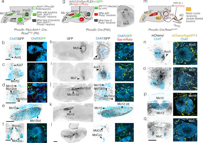
Fig. 3. Projections of IRtPhox2b and Peri5Atoh1 on hindbrain motoneurons.
a Strategy for the transgenic labeling of projections from Peri5Atoh1 (and RTN) and summary of the results; b–f Coronal (b–d, f) or parasagittal (e) sections through a P8 hindbrain showing GFP-labeled boutons (black) on motoneurons (blue) at medium (left) and high (right) magnification. g Strategy for the viral tracing of projections from IRtPhox2b and summary of the results (left), and mGFP-labeled infected cells of IRtPhox2b (right); h–l Coronal (h–j, l) or parasagittal (k) sections through a P56 hindbrain showing the GFP-labeled fibers (black) of IRtPhox2b neurons at low (left) and medium (middle) magnifications, and in extreme close-ups (right), together with Syp-mRuby labeled boutons (yellow) on motoneurons (blue). m schematic for retrograde tracing of premotor neurons for the right posterior digastric muscle, in a Phox2b::Cre;RosaSypGFP, and summary of the results. n–q (left) Coronal sections through the hindbrain at P8 showing the mCherry+ projections (black) of premotor neurons on the motor nuclei (ChAT+, blue); (right) close-ups on motoneurons receiving double-labeled Syp-GFP/mCherry boutons (yellow). Scale bars, b–f 200 μm for the left column, h–l 500 μm for the left column, n–q 200 μm for the left column.

