Figure 2.
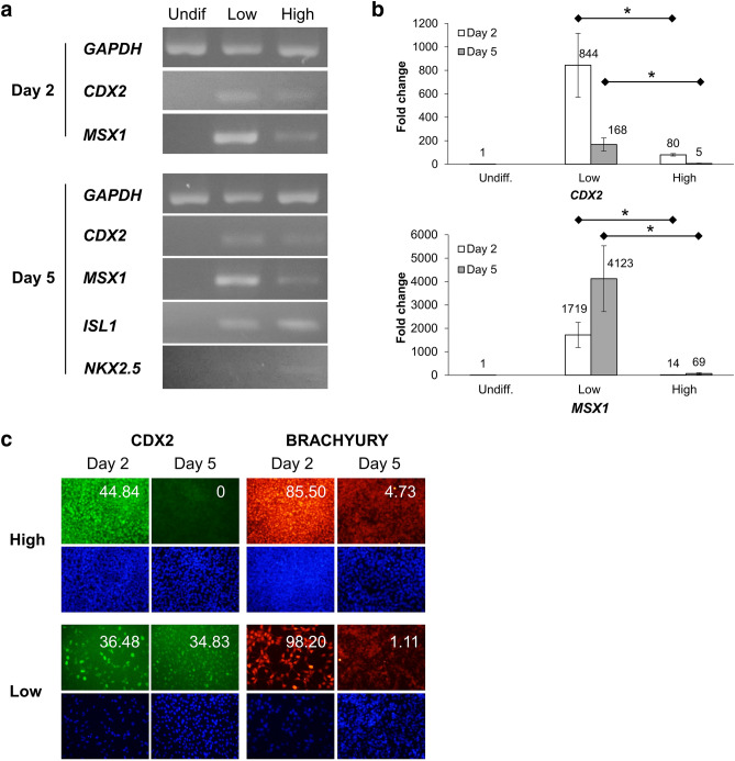
Cells at an initial low cell density expressed a higher level of anti-cardiac mesoderm genes, thereby inhibiting cardiac progenitor expression. (a) Expression of anti-cardiac (CDX2 and MSX1) and cardiac progenitor genes (ISL1 and NKX2.5) in undifferentiated cells and in cardiac-differentiated cells at initial low and high densities, on day 2 and 5 by RT-PCR. (b) Quantification of anti-cardiac gene expression (CDX2 and MSX1) on day 2 and 5 by RT-qPCR, showing the fold change in comparison with their expression in undifferentiated cells. Mean ± SE, * P < 0.05, t-tests with Holm’s correction, n = 2. (c) Immunostaining of cardiac differentiated cells at an initial low and high cell density using CDX2 (green) and BRACHYURY (red) on day 2 and day 5. DAPI (blue) was used for positive control. At an initial low cell density, CDX2 expression was not downregulated but was maintained at high levels on day 5. The numbers indicate the expression of the corresponding markers. Scale bar = 200 μm.

